Article

Secondary Highly Pathogenic Porcine Reproductive and Respiratory Syndrome Virus (HP-PRRSV2) Infection Augments Inflammatory Responses, Clinical Outcomes, and Pathogen Load in Glaesserella-parasuis-Infected Piglets

1
Shanghai Veterinary Research Institute, Chinese Academy of Agricultural Science, No. 518, Ziyue Road, Shanghai 200241, China
2
College of Agriculture, Hubei Three Gorges Polytechnic, Yichang 443000, China
*
Authors to whom correspondence should be addressed.
These authors contributed equally to this work.
Vet. Sci. 2023, 10(5), 365; https://doi.org/10.3390/vetsci10050365
Submission received: 14 March 2023 / Revised: 23 April 2023 / Accepted: 24 April 2023 / Published: 20 May 2023

Abstract

:

Simple Summary

Based on the fact that Gps are rooted in the upper respiratory tract of pigs, in order to investigate whether secondary infection with HP-PRRSV can exacerbate lung disease and chronic inflammation, our study was designed as follows. Our study randomly divided piglets into four groups: Gps + HP-PRRSV, Gps, HP-PRRSV, and controls. Piglets in the Gps + HP-PRRSV and Gps groups were infected through the intranasal route with the Gps W2 strain. The Gps + HP-PRRSV and HP-PRRSV groups were challenged with the HP-PRRSV HuN4 strain by intramuscular injection and intranasally at 5 days after the initial exposure to Gps. Alternatively, the control group animals received phosphate-buffered saline (PBS). Then, we observed the influence of HP-PRRSV–Gps coinfection on clinical outcomes, pathogen shedding and loading, cytokine production, and specific antibody levels at various time points in infected piglets. Our results revealed synergistic effects in HP-PRRSV–Gps coinfection, which increase the severity of clinical signs compared with single infections. Therefore, in the unavoidable situation of Gps infection in piglets, necessary measures must be made to prevent and control secondary infection of HP-PRRSV, which can save huge economic losses to the pork industry.

Abstract

Glaesserella parasuis (Gps), Gram-negative bacteria, are a universal respiratory-disease-causing pathogen in swine that colonize the upper respiratory tract. Highly Pathogenic Porcine Reproductive and Respiratory Syndrome Virus (HP-PRRSV2HP-PRRSV2) and Gps coinfections are epidemics in China, but little is known about the influence of concurrent coinfection on disease severity and inflammatory responses. Herein, we studied the effects of secondary HP-PRRS infection on clinical symptoms, pathological changes, pathogen load, and inflammatory response of Gps coinfection in the upper respiratory tract of piglets. All coinfected piglets (HP-PRRSV2 + Gps) displayed fever and severe lesions in the lungs, while fever was present in only a few animals with a single infection (HP-PRRSV2 or Gps). Additionally, HP-PRRSV2 and Gps loading in nasal swabs and blood and lung tissue samples was significantly increased in the coinfected group. Necropsy data showed that coinfected piglets suffered from severe lung damage and had significantly higher antibody titers of HP-PRRSV2 or Gps than single-infected piglets. Moreover, the serum and lung concentrations of inflammatory cytokines (TNF-α, IL-1β, IL-6, and IL-8) were also significantly higher in coinfected piglets than in those infected with HP-PRRSV2 or Gps alone. In conclusion, our results show that HP-PRRSV2 promotes the shedding and replication of Gps, and their coinfection in the upper respiratory tract aggravates the clinical symptoms and inflammatory responses, causing lung damage. Therefore, in the unavoidable situation of Gps infection in piglets, necessary measures must be made to prevent and control secondary infection with HP-PRRSV2, which can save huge economic losses to the pork industry.

1. Introduction

China is the largest pork-producing country [1]. Porcine Respiratory Disease Complex (PRDC) is the most challenging health concern for pig production worldwide, including in China [2]. PRDC often involves coinfections of viruses, such as swine Influenzas A Virus (swIAV) [3], PRRSV, and Porcine Circo Virus type 2 (PCV2), with bacteria such as Actinobacillus pleuropneumonia (APP), Mycoplasma hyopneumoniae (Mh), and Bordetella bronchiseptica (Bb) [4]. PRRSV can cause coinfection or secondary infection with bacteria such as Gps, Streptococcus suis (SS), and APP [3,5,6,7]. Notably, PRRSV outbreaks in July 2006 caused huge economic losses to the pork industry in China [8,9,10]. Such outbreaks can cause 100% morbidity and >40% mortality in infected piglets [11,12]. In PRRSV-infected animals, the respiratory tract becomes more prone to other pathogens, such as PCV2 [13,14,15], Classical swine fever virus (CSFV), SS, Gps, Mh, and various Salmonella species (spp), which may affect the severity of PRRSV-induced pathogenesis [16,17,18,19,20,21]. Previous studies on bacterial ecology in Chinese pig farms identified SS (77.92%), Gps (51.25%), Escherichia coli (52.39%), and Pasteurella multicide (26.77%) infections, along with the endemic of PRRSV, under field conditions [1,20,22]. Interestingly, studies comparing pathogens in healthy pigs with those with pneumonia found that PRRSV, PCV2, and Gps were prevalent among all samples regardless of the presence/absence of related clinical symptoms [23,24,25]. Overall, these data indicated that PRRSV and bacterial coinfections have high occurrences in the Chinese pig population [7]. In most cases, multiple infectious agents involved in the development of clinical conditions make the universal reductionist approach impractical to examine the host–pathogen interactions, as in the case of single infections [20].
Gps is a Gram-negative, non-hemolytic, nicotinamide adenine dinucleotide (NAD) dependent bacterium [26]. Gps is not only a universal bacterium of the upper respiratory tract of pigs, but also an important universal respiratory pathogen causing fibrinous polyserositis, arthritis, and meningitis in pigs [27,28,29,30]. Gps infections lead to high mortality rates in pigs, causing significant economic losses to the swine industry worldwide [31,32]. Gps strain can also be obtained from the nasal cavities, tonsils, and tracheae of healthy pigs [33,34]. Under favorable conditions, Gps activates innate immune responses promoting the production of inflammatory cytokines [35]. Interestingly, PRRSV infection can have an additive effect on Gps infection and loading [33]. However, the factors causing Gps systemic infection in pigs have not yet been identified [36].
The PRRSV is a single-stranded positive-sense RNA virus with a capsid [37,38,39]. The PRRSV disease was first identified in the United State of America in the late 1980s and in Germany in the 1990s [40,41,42,43,44], and then it spread to other pig-producing areas, including China, causing enormous economic losses to the pig industry [45,46,47,48]. PRRSV is classified in the order Nidovirales, genus Arterivirus, family Arteviridae, together with Equine Arteritis Virus (EAV), mouse Lactate Dehydrogenase-elevating Virus (LDV), and Simian Hemorrhagic Fever Virus (SHFV) [42,49,50]. Based on genetic and antigenic determinants, PRRSV is divided into two major genotypes: PRRSV1 (the European Lelystad strain) and PRRSV2 (the North American Vr-2332 strain) [51,52]. Phylogenetically, PRRSV1 is further classified into three subtypes [53,54]: pan-European subtype 1, and East-European subtypes 2 and 3, while PRRSV2 is classified into nine distinct subtypes [7,55]. PRRSV replicates in monocytic lineage cell types, particularly porcine alveolar macrophages (PAMs), and the viral disease is characterized by severe interstitial pneumonia, reduced growth, and high mortality in young piglets, and reproductive failure in sows [20,56]. In China, PRRSV2 was first reported by Baoqing Guo in 1996 [57], while HP-PRRSV2 was identified by Kegong Tian from the Jiangxi Province in 2007 [58]. Then, HP-PRRSV2 spread to other pig-producing regions, causing serious pecuniary losses. The NADC30-like PRRSV2 was described by Zhao in 2015 [14,59,60]. HP-PRRS2V and NADC30-like PRRSV2 became prevalent after 2015 [20]. PRRSV1 and NADC34-like PRRSV2 were separately reported by Nanhua Chen in 2011 [61] and Hongliang Zhang in 2018 [62], which increased the difficulty of prevention and control of PRRSV in China [20].
PRRSV infection makes pigs susceptible to secondary infection by damaging PAM cells and inducing nasal mucositis [63]. For instance, a high detection rate of Gps is common in pig farms infected with PRRSV [64]. Yu et al. found that HP-PRRSV2 promotes Gps proliferation in blood and tissues, which increases the susceptibility to Gps in PRRSV-positive pigs [31]. Li et al. found that the transfection of Gps RNA enhanced HP-PRRSV2-mediated inflammatory response in coinfected animals [7]. Zhang et al. showed that the PRRSV2–Gps coinfection aggravated lung diseases and chronic inflammation by modulating host gene expression [33]. In the current trend, the high recombination rate and spread ability of PRRSV strains increase the incidence and mortality rates in Gps infections [65].
Although HP-PRRSV2–Gps coinfection is very common, little is known about the effects of coinfection on the pathogenicity, disease severity, inflammatory mediators, and antibody responses in infected piglets [66]. Studying the mechanism of coinfection can help control Gps outbreaks. Accordingly, in this study, we investigated the influence of HP-PRRSV2–Gps coinfection on clinical outcomes, pathogen shedding and loading, cytokine production, and specific antibody levels at various time points in infected piglets. Our results revealed synergistic effects in HP-PRRSV2–Gps coinfection, which increase the severity of clinical signs compared with single infections.

2. Materials and Methods

2.1. Ethics Statements

All animal experiments were carried out according to ethical guidelines and were approved by the Institutional Laboratory Animal Care and Use Committee of Shanghai Veterinary Research Institute (SHVRI) CAAS, Shanghai (IACUC no: SHVRI-P-2018010503), and performed in compliance with the Guidelines on Humane Treatment of Laboratory Animals (Ministry of Science and Technology of the People’s Republic of China, policy no. 2006 398).

2.2. Virus and Bacterium

The HP-PRRSV2 HuN4 strain (GenBank: EF635006) was kindly provided by Professor Guangzhi Tong (Shanghai Veterinary Research Institute, Chinese Academy of Agricultural Sciences). The virus was propagated in the MARC-145 cell line and cultured in Dulbecco’s modified eagle’s medium (DMEM; Invitrogen, Waltham, MA, USA), with 10% fetal bovine serum (FBS; Thermo Scientific, Waltham, MA, USA) and antibiotics (100 units of penicillin, 10 mg streptomycin, and 25 mg amphotericin B per mL; Sigma-Aldrich, St. Louis, MO, USA) at 37 °C and 5% CO2. The stock virus titer for experimental infection was 3 × 10³ TCID50/mL [45].
The virulent Gps W2 strain (serotype 13) was isolated from Shanghai, China, in March 2016, and confirmed by agglutination and agar diffusion tests. Gps was cultured on tryptic soy agar (TSA; Difco Laboratories, Franklin Lakes, NJ, USA) containing 10 mg/mL NAD and 5% calf bovine serum. The amount of W2 strain used for intranasal infection was 3 × 10⁸ CFU/mL [67].

2.3. Pigs, Experimental Design, and Sampling

Thirty-two 4-week-old castrated male DLY (Duroc  ×  Landrace  ×  Yorkshire) weaned piglets from the same commercial farm were used in this study. Before beginning the experiments, all piglets were tested for PRRSV, CSFV, PCV2, swine influenza virus (SIV), and Gps antigen by quantitative real-time PCR (qRT-PCR), and for PRRSV and Gps antibodies using commercial ELISA kits. All piglets were negative for PRRSV and Gps. After arrival at the animal house, the animals were acclimated for 7 days before starting the experiment.
Piglets were stochastically divided into four groups (n = 8): Gps + HP-PRRSV2, Gps, HP-PRRSV2, and control. Each group was housed in a separate room and provided with a basal diet (based on the NRC (2012) recommendation for the nutrient requirements of weaned piglets) [68] and water throughout the experiment.
Piglets in the Gps+ HP-PRRSV2 and Gps groups were infected through the intranasal route with 2.0 mL of Gps W2 strain (3 × 10⁸ CFU/mL). The Gps + HP-PRRSV2 and HP-PRRSV2 groups were challenged with 3.0 mL of HP-PRRSV2 HuN4 strain (3 × 10³ TCID50) by intramuscular injection (1 mL) and intranasally (2 mL) at 5 days after the initial exposure to Gps. Alternatively, the control group animals received phosphate-buffered saline (PBS). The dose and timing of HP-PRRSV2 inoculation were according to Yang et al. (2012).
Piglets were monitored daily for clinical signs, and rectal temperatures were recorded before feeding. At 0, 3, 6, 10, 14, 17 and 21 days post-infection (dpi), nasal swabs and blood samples were collected to determine the pathogen (HP-PRRSV2 /Gps) load, HP-PRRSV2/Gps antibody titer, and the serum levels of inflammatory cytokines. Three pigs per group were randomly euthanized at 6 days post-HP-PRRSV2 challenge, and blood, heart, liver, spleen, lung, and kidney samples were collected for analysis. The remaining pigs in each group were euthanized at 21 dpi.

2.4. Clinical and Pathological Evaluation

After Gps and HP-PRRSV2 challenge, piglets were monitored daily and scored for clinical signs, including mental state, rectal temperature, anorexia, dyspnea, coughing, and other symptoms, such as mortality, trembling, cyanosis, vomiting, diarrhea, and limping.
The piglets were scored according to the scoring criteria in Table 1. All scores were accumulated to give a total clinical score for each pig (0–8) [67]. For necropsy, lung sections were fixed in 10% neutrally buffered formalin for histological examination using hematoxylin and eosin (H&E) staining (Wuhan Service Biotechnology Co. Ltd., Wuhan, China), and the histopathological changes were observed under an optical microscope (Olympus, Tokyo, Japan).

2.5. Detection of HP-PRRSV2/Gps Antibodies by ELISA

Serum samples were detected by an enzyme-linked immunosorbent assay (ELISA) kit for PRRSV antibodies (IDEXX Laboratories, Atlanta, GA, USA) and a swine Gps ELISA kit (Biovet, Karnataka, India) for Gps antibodies, according to the manufacturers’ instructions.

2.6. Detection of Pathogen Load by qRT-PCR

Bacterial DNA was extracted using the DNeasy Blood and Tissue Kit (QIAGEN, Tokyo, Japan) and viral RNA was extracted with a Viral DNA/RNA Kit (OMEGA, Tokyo, Japan), according to the manufacturers’ instructions [67]. The quantity of Gps and HP-PRRSV2 in piglet samples was detected by qRT-PCR [31,44] performed on an ABI Step One thermocycler (Applied Biosystem, Waltham, MA, USA).

2.7. Cytokine Assays

Piglet lung tissues from necropsy were analyzed for cytokines [67]. Briefly, 1 g of lung tissue was homogenized in 1 mL of PBS (1:1, w/v) with an X620 tissue homogenizer (CAT). The homogenate was centrifuged at 3000 g for 10 min and the obtained supernatants were stored at −80 °C for cytokine analysis. Piglet serum samples were collected at 0, 3, 6, 7, 14, 17, and 21 dpi. The lung tissues and serum concentrations of inflammatory factors (IL-1β, TNF-α, IL-6, and IL-8) were quantified with respective ELISA kits (R&D Systems, Minneapolis, MN, USA), following standard protocols. The corresponding mRNA levels were determined using an ABI Step One thermocycler (Applied Biosystems) and relative gene expression levels were normalized against β-actin using the 2−ΔΔCT method [69]. All tests were performed in triplicate.

2.8. Statistical Analysis

All statistical analyses were performed using Student’s t-tests in SPSS version 16.0 (SPSS Inc, Chicago, IL, USA). Data are presented as the mean ± standard deviation (SD), and those with p-value < 0.05 were considered statistically significant.

3. Results

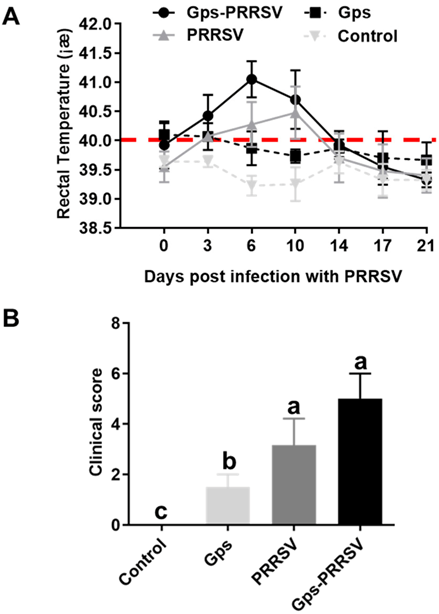
3.1. Clinical Evaluation

Piglets challenged with Gps alone showed mild clinical signs, while the same was absent in the control group throughout the experiment. However, the animals infected with HP-PRRSV2 alone or coinfected with Gps developed significant clinical signs including depression, drowsiness, loss of appetite, fever, anorexia, lethargy, coughing, shivering, and breathing difficulties. Among the two, the HP-PRRSV2–Gps-coinfected group was more severely affected than the HP-PRRSV2 alone group (Figure 1).
Animals inoculated with Gps alone exhibited low-grade fever at 1 and 2 dpi (temperature 40–40.5 °C), and only 50% of the animals (3/6) displayed a rise in body temperature. On the other hand, piglets in the Gps + HP-PRRSV2 and HP-PRRSV2 alone groups had fever following HP-PRRSV2 infection (rectal temperature ≥ 40 °C). Moreover, high rectal temperatures were observed in piglets exposed to HP-PRRSV2 alone from 3 to 12 dpi (Figure 1A).
HP-PRRSV2–Gps coinfection triggered major responses: the mean rectal temperature was 41 °C at 6 days after the HP-PRRSV2 challenge and remained higher than normal until 14 dpi (Figure 1A). Animals exposed to Gps alone did not show any significant rise in body temperature compared with the control group. There were significant differences among mean rectal temperatures in animals of the three infected groups. However, at 14 days after HP-PRRSV2 infection, fever disappeared in all challenged animals (Figure 1A). Clinical scores in animals were obtained by observing clinical symptoms (dyspnea, coughing, runny nose, anorexia, limping, and diarrhea). These clinical symptoms were most apparent from day 3 to day 10 in the HP-PRRSV2–Gps-coinfected group. The mean clinical scores (±SD) in all groups are shown in Figure 1B. Individual clinical scores ranged from 0 to 2 in Gps alone (1.50 ± 0.41), and 0 to 4 in the HP-PRRSV2 alone group (3.17 ± 0.73), while the same ranged from 2 to 6 in coinfected pigs (5.12 ± 0.61) (Figure 1B). Pigs in the control group did not exhibit any clinical signs. There were statistically significant differences in the mean clinical scores among the three infected groups (p < 0.05).

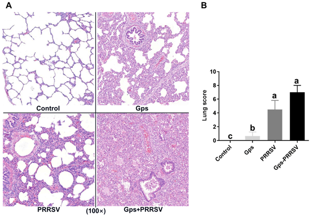
3.2. Pathological Examination

We conducted post-mortem examinations at 6 dpi and the results revealed gross lesions, including lung congestion or consolidation, inguinal lymph node tumidity and hemorrhage, submandibular lymph node tumidity and hemorrhage, liver congestion tumidity, spleen infarction, kidney hemorrhage or grey spot, and brain hemorrhage or edema in the coinfected group (Figure 2A). The HP-PRRSV2 alone group also displayed pulmonary interstitial pneumonia and pulmonary alveolar epithelial damage, but these viral-associated lesions were mild compared with coinfected piglets (Figure 2A). Three of the coinfected animals presented serositis (3/6), while serositis was completely absent in the Gps alone group. All control animals remained healthy throughout the experiment.
Lung scores were assigned according to the pathological degree of infected lung tissue. The mean lung scores (±SD) are shown in Figure 2B. There were significant statistical differences between the coinfected group and the Gps alone group (0.75 ± 0.35) (p < 0.05), while animals challenged with HP-PRRSV2 alone (4.50 ± 0.78) exhibited lung damage similar to that of the coinfected group (7.05 ± 0.61). However, the damage was more serious in coinfection with a significantly greater abnormal area than the HP-PRRSV2 infection alone (Figure 2A). Furthermore, there were no significant differences among the Gps and control groups, whereas both the Gps- and HP-PRRSV2-alone-infected animals exhibited significant differences in lung scores (p < 0.05; Figure 2B).

3.3. Pathogen Shedding and Load

Coinfected pigs had higher Gps and HP-PRRSV2 titters in nasal swabs (Figure 3A,D). In coinfected piglets, the mean Gps titers in nasal swabs were significantly higher from 3 dpi and remained elevated until the end of the study (Figure 3A; p < 0.01). The load of HP-PRRSV2 in nasal swabs in coinfected piglets reached a maximum value on day 6 after HP-PRRSV2 infection, and the difference from the HP-PRRSV2 single-infection group was statistically significant (p < 0.05). At day 10 after HP-PRRSV2 infection, the load of HP-PRRSV2 in nasal swabs from the coinfected and HP-PRRSV2 single-infected groups decreased abruptly, with no statistical differences between the two groups (Figure 3D; p > 0.05).
The blood titers of Gps and HP-PRRSV2 are shown in Figure 3B,E. Gps was non-detectable in the Gps alone group, but it appeared in coinfected animals at 6 days after HP-PRRSV2 infection (peaked at 14 dpi), showing a significant difference (p < 0.01) from single-infected animals (Figure 3B). However, HP-PRRSV2 was detected in serum samples from both coinfected and HP-PRRSV2-alone-infected piglets at 3 days post-challenge (Figure 3E). The copy number of HP-PRRSV2 reached the maximum at 6 dpi in both groups, which was significantly higher (p < 0.05) in coinfected animals (Figure 3E).
Lung tissue samples were collected at 6 dpi to further determine the Gps and HP-PRRSV2 titers in single- and coinfected animals. The results were similar to those obtained for serum samples: Gps was undetectable in Gps single-infected animals, whereas coinfected animals had significantly higher mean genomic copies of Gps (Figure 3C; p < 0.01). HP-PRRSV2 was detected at a relatively high copy number in lung tissues from HP-PRRSV2-alone-infected piglets compared with coinfected animals (Figure 3F; p < 0.05). Together, these results are consistent with previous studies showing that Gps infection enhances HP-PRRSV2 replication [31]. This also validates our hypothesis that coinfection accelerates both HP-PRRSV2 and Gps replication in coinfected animals, causing greater damage than a single infection.

3.4. Serum Antibody Levels of Gps and HP-PRRSV2

Specific humoral immune response against HP-PRRSV2 and Gps in challenged animals is presented in Figure 4. At 0, 6, 14, and 21 days after HP-PRRSV2 infection, serum samples were determined for antibody levels by ELISA. The coinfected piglets produced specific antibodies against Gps, while animals infected with Gps alone did not exhibit a significant humoral response (Figure 4A). At 14 dpi, the coinfected animals produced much higher levels of Gps antibodies, which remained high (p < 0.05) until the end of the experiment (21 dpi; Figure 4A). By contrast, HP-PRRSV2 triggered antibody levels showed similar patterns in coinfected and HP-PRRSV2 alone groups (Figure 4B); however, S/P levels in coinfected animals were significantly higher than in HP-PRRSV2 single-infected animals at 14 and 21 dpi (Figure 4B). Consistent with a previous study in 2012 [31], these findings confirmed that HP-PRRSV2 infection enhanced Gps replication in coinfected animals, which promoted a strong antibody response against Gps.

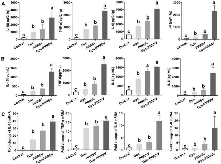
3.5. Cytokine Analysis

Microbial infection can change cytokine levels in animals [3,67]. Herein, we measured changes in the expression and abundance of cytokines TNF-ɑ, IL-1β, IL-8, and IL-6 at 6 dpi in lung tissue homogenates and serum by ELISA and qRT-PCR [70]. The levels of IL-1β, TNF-ɑ, and IL-8 were significantly higher (p < 0.05) in coinfected animals (up to 1500, 3000, and 3000 pg/mL, respectively), while both the single-infected animals had similar titers (p > 0.05; Figure 5A,B). Similar patterns were observed for TNF-ɑ and IL-6 in lung and serum samples. However, the IL-6 serum titer did not significantly differ between the HP-PRRSV2 single-infected and coinfected animals (Figure 5A,B).
Furthermore, there were no statistical differences in gene expression levels of IL-1β and TNF-ɑ among both types of single-challenged animals (Figure 5C), while IL-1β and TNF-α were upregulated in co-infected animals. In addition, the expression levels of IL-8 and IL-6 were much lower in both types of single-infected animals (Figure 5C). Interestingly, lung tissues from coinfected animals displayed an upregulation of IL-6 (12-fold) and IL-8 (14-fold) more than the single-infected groups (Figure 5C). The tested cytokines were statistically more upregulated in all infected animals than in control animals (Figure 5).
Concisely, in general, the maximum levels of cytokines were observed in coinfected animals at 6 dpi (Figure 5). Gps alone infection did not upregulate any of the investigated cytokines, while HP-PRRSV2 exposure following Gps inoculation stimulated the production of the same nature. These results are consistent with the localization of gross lung lesions: significant correlations were observed between pathological changes in the lungs and concentrations of TNF-ɑ, IL-1β, IL-8, and IL-6.

4. Discussion

Although single infections with bacteria or viruses alone can induce respiratory infections in pigs, coinfection with various pathogens is very common [28,71]. A Gps infection initiates an innate immune response and induces the production of inflammatory cytokines [72,73]. HP-PRRSV2 targets the immune system of pigs, impairing immune defense against pathogenic microbes and increasing host susceptibility to primary and secondary pathogens [64,74]. In this study, we found that HP-PRRSV2–Gps coinfection had aggravated clinical outcomes, increased pathogen shedding and load, and specific antibody and cytokine production at various time points following HP-PRRSV2 challenge after Gps infection. We observed that the coinfection enhanced the severity of the disease in different ways, including increased Gps and HP-PRRSV2 replication, which modulated the inflammatory response. In addition, pulmonary lesions in coinfected animals were more severe compared to those in single-infected animals.
Previous studies reported that piglets infected with HP-PRRSV2 alone or in combination with Gps suffered from severe interstitial pneumonia [7,74,75]. It is also suggested that secondary bacterial infection followed by HP-PRRSV2 infection exacerbates illness and mortality in infected piglets [76,77,78]. Here, we provided more comprehensive information on the effect of HP-PRRSV2 and Gps coinfection on respiratory disease pathogenicity in challenged piglets. We examined Gps and HP-PRRSV2 mRNA levels in nasal swabs, blood, and lung tissues at various time points of coinfection and single infection. The Gps load in nasal swabs was significantly higher from day 3 to 17 in coinfected animals compared with Gps single-infected animals (Figure 3A). Additionally, the serum Gps loads were significantly different between the coinfected and Gps single-infected animals at 10 and 14 days post-HP-PRRSV2 infection (Figure 3B). There was a significant increase in the Gps copy number in lung tissues of coinfected animals, while Gps was undetectable in Gps single-infected pigs (Figure 3C). From the above results, we concluded that HP-PRRSV2 infection promotes the proliferation of Gps in the nose, blood, and lungs.
Based on our in vivo findings, we concluded that Gps alone infection did not cause significant lesions in the lungs (Figure 2). Clinical signs and symptoms showed no rise in temperature, and even the post-mortem examination confirmed the same. This indicated that PPRSV2 infection enhances Gps colonization, which ultimately increases its copy number in the lung and blood (Figure 3). These in vivo findings are consistent with a previous report showing that Gps causes typical systemic polyserositis lesions only after coinfection with HP-PRRSV2 [78,79]. Notably, Mycoplasma hyrhinis coinfection with HP-PRRSV2 can also enhance pathological lesions in the lungs [80], which is consistent with our results showing that Gps coinfection with HP-PRRSV2 leads to lung consolidation and severe interstitial pneumonia.
We further investigated whether Gps infection affects HP-PRRSV2 pathogenicity. HP-PRRSV2 proliferation in nasal mucosa takes a very short time. There were significant differences of PRRSV load in the nasal mucosa between coinfection and HP-PRRSV2 single-infection groups at 6 dpi (Figure 3A). Moreover, HP-PRRSV2 copy numbers (400) were significantly higher in the coinfected animal blood and lung tissues (Figure 3B,C). From the above results, we concluded that Gps infection has a significant impact on HP-PRRSV2 pathogenicity. Additionally, lung consolidation and severe interstitial pneumonia were observed in coinfected animals (Figure 2). Enhanced HP-PRRSV2 replication in coinfected animals led to more severe clinical outcomes, involving earlier and greater immune and inflammatory responses (Figure 4 and Figure 5). This conclusion is consistent with the study of Yu et al. in 2012 [31].
How the coinfection of viruses and bacteria enhances disease severity is not fully understood [81]. Coinfection often upregulates various cytokines that help microbes with replication, but causes detrimental damage to infected tissues [3,82]. HP-PRRSV2 infection has several mechanisms that make infected animals prone to secondary bacterial infection, including increased expression of cellular receptors that enhance colonization and modification of host immune responses [7,83,84]. Here, we found that the production of inflammatory cytokines (TNF-ɑ, IL-1β, IL-8, and IL-6) was significantly increased in coinfected animals compared with both types of single-infected pigs (Figure 5). This is consistent with a previous study showing that PRRSV infection upregulated cytokines (TNF-ɑ, IL-1β, IL-8, and IL-6) in pig sera and promoted the bacterial load of 11 bacterial species in the lungs, including Gps (Li et al., 2017). In this study, coinfection upregulated inflammatory cytokine in lung tissue and blood as well (Figure 5), but the exact molecular mechanism is unknown.
HP-PRRSV2 and Gps are known to stimulate each other’s replication when coinfected, but the impacts of coinfection on clinical outcomes, the kinetics of the immune system and inflammatory responses, and pathogen load and shedding in piglets remain poorly understood [7,31]. Herein, piglets challenged with HP-PRRSV2 alone displayed significant differences in cytokine production, lung score, and macroscopic lesions in the lungs compared with coinfected animals (Figure 2 and Figure 5). The mRNA levels and antibody titer results showed that both HP-PRRSV2 and Gps replicated more rapidly and elicited more severe local inflammatory responses in coinfected animals than in any of the single-infected animals (Figure 3 and Figure 4). A similar trend has been reported for SIV and Gps coinfection [67]. The significant influence of both pathogens on the systemic inflammatory response was only present in coinfected animals (Figure 5). These outcomes are consistent with the fact that PRRSV and S. suis serotype 2 coinfection upregulated TNF-α, IL-1β, IL-8, and IL-6 gene expressions, compared with single-infected animals [7]. Nonetheless, further studies are needed to elucidate the molecular mechanism by which HP-PRRSV2–Gps coinfection enhances the production of pro-inflammatory cytokines increasing the disease severity.

5. Conclusions

There were lung lesions among all of the challenged piglets. HP-PRRSV2 infection potentiates the degree of lung lesions and facilitates Gps replication in porcine lungs. Gps infection enhances the copy number of HP-PRRSV2 in nasal shedding, blood, and lung tissues. Enhanced Gps and HP-PRRSV2 replication and stronger systemic/local inflammatory responses aggravate clinical signs in coinfected piglets. The coinfected piglets had a more severe fever, pathological changes, microscopic lung lesions, and a higher pathogen load than any of the single-infected piglets. These results showed that the secondary infection of HP-PRRSV2 will aggravate lung disease and chronic inflammation in the case of common Gps infection of the upper respiratory tract in piglets. Therefore, it is necessary to prevent and control HP-PRRSV2 infection in such cases.

Author Contributions

Conceptualization, J.W.; Methodology, Y.O., J.Z. and J.W.; Software, B.L.; Validation, Y.J. and Y.Q.; Formal analysis, Z.L.; Investigation, K.L.; Resources, Z.M.; Writing—original draft, L.P.; Writing—review & editing, Z.G. and J.W.; Visualization, D.S.; Supervision, Z.M. All authors have read and agreed to the published version of the manuscript.

Funding

This work was supported by the National Natural Science Foundation of China (no. 31972693 awarded to Y.Q.), the Shanghai Agriculture Applied Technology Development Program, China (no. X2022-02-08-00-12-F01195 and no. X2021-02-08-00-12-F00770 awarded to J.W.), and the National Basic Research Program (973 Plan) (no. 2014CB542703 awarded to J.W.).

Institutional Review Board Statement

Piglets included in this survey were castrated male DLY (Duroc × Landrace × Yorkshire) weaned piglets kept in a separate room in accordance with NRC (2012) recommendation for the nutrient requirements of weaned piglets. All animal experiments were approved by the Institutional Animal Care and Use Committee of Shanghai Veterinary Research Institute, China (IACUC No: SVRI-P-2020101206) and performed in compliance with the Guidelines on the Hu-mane Treatment of Laboratory Animals (Ministry of Science and Technology of the People’s Republic of China, Policy No. 2006 398).

Informed Consent Statement

Not applicable.

Data Availability Statement

Not applicable.

Acknowledgments

Not applicable.

Conflicts of Interest

The authors declare no conflict of interest.

References

  1. Zhu, H.; Chang, X.; Zhou, J.; Wang, D.; Zhou, J.; Fan, B.; Ni, Y.; Yin, J.; Lv, L.; Zhao, Y.; et al. Co-infection analysis of bacterial and viral respiratory pathogens from clinically healthy swine in Eastern China. Vet. Med. Sci. 2021, 7, 1815–1819. [Google Scholar] [CrossRef] [PubMed]
  2. Du, S.; Xu, F.; Lin, Y.; Wang, Y.; Zhang, Y.; Su, K.; Li, T.; Li, H.; Song, Q. Detection of Porcine Circovirus Type 2a and Pasteurella multocida Capsular Serotype D in Growing Pigs Suffering from Respiratory Disease. Vet Sci. 2022, 9, 528. [Google Scholar] [CrossRef] [PubMed]
  3. Auray, G.; Lachance, C.; Wang, Y.; Gagnon, C.A.; Segura, M.; Gottschalk, M. Gottschalk. Transcriptional Analysis of PRRSV-Infected Porcine Dendritic Cell Response to Streptococcus suis Infection Reveals Up-Regulation of Inflammatory-Related Genes Expression. PLoS ONE 2016, 11, e0156019. [Google Scholar] [CrossRef] [PubMed]
  4. Tonni, M.; Formenti, N.; Boniotti, M.B.; Guarneri, F.; Scali, F.; Romeo, C.; Pasquali, P.; Pieters, M.; Maes, D.; Alborali, G.L. The role of co-infections in M. hyopneumoniae outbreaks among heavy fattening pigs: A field study. Vet. Res. 2022, 53, 41. [Google Scholar] [CrossRef]
  5. Saade, G.; Deblanc, C.; Bougon, J.; Marois-Créhan, C.; Fablet, C.; Auray, G.; Belloc, C.; Leblanc-Maridor, M.; Gagnon, C.A.; Zhu, J.; et al. Coinfections and their molecular consequences in the porcine respiratory tract. Vet. Res. 2020, 51, 80. [Google Scholar] [CrossRef]
  6. Ouyang, T.; Zhang, X.; Liu, X.; Ren, L. Co-Infection of Swine with Porcine Circovirus Type 2 and Other Swine Viruses. Viruses 2019, 11, 185. [Google Scholar] [CrossRef]
  7. Li, J.; Wang, S.; Li, C.; Wang, C.; Liu, Y.; Wang, G.; He, X.; Hu, L.; Liu, Y.; Cui, M.; et al. Secondary Haemophilus parasuis infection enhances highly pathogenic porcine reproductive and respiratory syndrome virus (HP-PRRSV) infection-mediated inflammatory responses. Vet. Microbiol. 2017, 204, 35–42. [Google Scholar] [CrossRef]
  8. Chen, N.; Xiao, Y.; Ye, M.; Li, X.; Li, S.; Xie, N.; Wei, Y.; Wang, J.; Zhu, J. High genetic diversity of Chinese porcine reproductive and respiratory syndrome viruses from 2016 to 2019. Res. Vet. Sci. 2020, 131, 38–42. [Google Scholar] [CrossRef]
  9. Dunkelberger, J.R.; Serão, N.V.L.; Weng, Z.; Waide, E.H.; Niederwerder, M.C.; Kerrigan, M.A.; Lunney, J.K.; Rowland, R.R.R.; Dekkers, J.C.M. Genomic regions associated with host response to porcine reproductive and respiratory syndrome vaccination and co-infection in nursery pigs. BMC Genom. 2017, 18, 865. [Google Scholar] [CrossRef]
  10. Brockmeier, S.L.; Loving, C.L.; Palmer, M.V.; Spear, A.; Nicholson, T.L.; Faaberg, K.S.; Lager, K.M. Comparison of Asian porcine high fever disease isolates of porcine reproductive and respiratory syndrome virus to United States isolates for their ability to cause disease and secondary bacterial infection in swine. Vet. Microbiol. 2017, 203, 6–17. [Google Scholar] [CrossRef]
  11. Han, J.; Zhou, L.; Ge, X.; Guo, X.; Yang, H. Pathogenesis and control of the Chinese highly pathogenic porcine reproductive and respiratory syndrome virus. Vet. Microbiol. 2017, 209, 30–47. [Google Scholar] [CrossRef]
  12. Zhou, H.; Pan, Y.; Liu, M.; Han, Z. Prevalence of Porcine Pseudorabies Virus and Its Coinfection Rate in Heilongjiang Province in China from 2013 to 2018. Viral Immunol. 2020, 33, 550–554. [Google Scholar] [CrossRef]
  13. Chung, W.B.; Chan, W.H.; Chaung, H.C.; Lien, Y.; Wu, C.C.; Huang, Y.L. Real-time PCR for quantitation of porcine reproductive and respiratory syndrome virus and porcine circovirus type 2 in naturally-infected and challenged pigs. J. Virol. Methods 2005, 124, 11–19. [Google Scholar] [CrossRef]
  14. Zhang, J.; Wang, P.; Xie, C.; Ha, Z.; Shi, N.; Zhang, H.; Li, Z.; Han, J.; Xie, Y.; Qiu, X.; et al. Synergistic Pathogenicity by Coinfection and Sequential Infection with NADC30-like PRRSV and PCV2 in Post-Weaned Pigs. Viruses 2022, 14, 193. [Google Scholar] [CrossRef]
  15. Tregaskis, P.L.; Staines, A.; Gordon, A.; Sheridan, P.; McMenamy, M.; Duffy, C.; Collins, P.J.; Mooney, M.H.; Lemon, K. Co-infection status of novel parvovirus’s (PPV2 to 4) with porcine circovirus 2 in porcine respiratory disease complex and porcine circovirus-associated disease from 1997 to 2012. Transbound. Emerg. Dis. 2021, 68, 1979–1994. [Google Scholar] [CrossRef]
  16. Albina, E.; Mesplede, A.; Chenut, G.; Le Potier, M.F.; Bourbao, G.; Le Gal, S.; Leforban, Y. A serological survey on classical swine fever (CSF), Aujeszky’s disease (AD) and porcine reproductive and respiratory syndrome (PRRS) virus infections in French wild boars from 1991 to 1998. Vet. Microbiol. 2000, 77, 43–57. [Google Scholar] [CrossRef]
  17. Xu, M.; Wang, S.; Li, L.; Lei, L.; Liu, Y.; Shi, W.; Wu, J.; Li, L.; Rong, F.; Xu, M.; et al. Secondary infection with Streptococcus suis serotype 7 increases the virulence of highly pathogenic porcine reproductive and respiratory syndrome virus in pigs. Virol. J. 2010, 7, 184. [Google Scholar] [CrossRef]
  18. Palzer, A.; Eddicks, M.; Zoels, S.; Stark, J.; Reese, S.; Strutzberg-Minder, K.; Fiebig, K.; Ritzmann, M. Field evaluation of the efficacy, compatibility and serologic profiling of a combined vaccine against porcine reproductive and respiratory syndrome and Haemophilus parasuis in nursery pigs. Prev. Vet. Med. 2015, 119, 134–140. [Google Scholar] [CrossRef]
  19. Thacker, E.L.; Thacker, B.J.; Young, T.F.; Halbur, P.G. Effect of vaccination on the potentiation of porcine reproductive and respiratory syndrome virus (PRRSV)-induced pneumonia by Mycoplasma hyopneumoniae. Vaccine 2000, 18, 1244–1252. [Google Scholar] [CrossRef]
  20. Zhao, D.; Yang, B.; Yuan, X.; Shen, C.; Zhang, D.; Shi, X.; Zhang, T.; Cui, H.; Yang, J.; Chen, X.; et al. Advanced Research in Porcine Reproductive and Respiratory Syndrome Virus Co-infection with Other Pathogens in Swine. Front. Vet. Sci. 2021, 8, 699561. [Google Scholar] [CrossRef]
  21. Zuckermann, F.A.; Husmann, R.; Chen, W.; Roady, P.; Pfeiff, J.; Leistikow, K.R.; Duersteler, M.; Son, S.; King, M.R.; Augspurger, N.R. Bacillus-Based Direct-Fed Microbial Reduces the Pathogenic Synergy of a Coinfection with Salmonella enterica Serovar Choleraesuis and Porcine Reproductive and Respiratory Syndrome Virus. Infect. Immun. 2022, 90, e0057421. [Google Scholar] [CrossRef] [PubMed]
  22. Day, D.N.; Sparks, J.W.; Karriker, L.A.; Stalder, K.J.; Wulf, L.W.; Zhang, J.; Kinyon, J.M.; Stock, M.L.; Gehring, R.; Wang, C.; et al. Impact of an experimental PRRSV and Streptococcus suis coinfection on the pharmacokinetics of ceftiofur hydrochloride after intramuscular injection in pigs. J. Vet. Pharmacol. Ther. 2015, 38, 475–481. [Google Scholar] [CrossRef] [PubMed]
  23. Nelsen, A.; Lin, C.-M.; Hause, B.M. Porcine Parvovirus 2 Is Predominantly Associated with Macrophages in Porcine Respiratory Disease Complex. Front. Vet. Sci. 2021, 8, 726884. [Google Scholar] [CrossRef] [PubMed]
  24. van Dixhoorn, I.D.E.; Beest, D.E.T.; Bolhuis, J.E.; Parmentier, H.K.; Kemp, B.; van Mourik, S.; Stockhofe-Zurwieden, N.; Van Reenen, C.G.; Rebel, J.M.J. Animal-Based Factors Prior to Infection Predict Histological Disease Outcome in Porcine Reproductive and Respiratory Syndrome Virus- and Actinobacillus pleuropneumoniae-Infected Pigs. Front. Vet. Sci. 2021, 8, 742877. [Google Scholar] [CrossRef]
  25. Yan, X.; Zeng, J.; Li, X.; Zhang, Z.; Din, A.U.; Zhao, K.; Zhou, Y. High incidence and characteristic of PRRSV and resistant bacterial Co-Infection in pig farms. Microb. Pathog. 2020, 149, 104536. [Google Scholar]
  26. Fu, S.; Xu, L.; Li, S.; Qiu, Y.; Liu, Y.; Wu, Z.; Ye, C.; Hou, Y.; Hu, C.A. Baicalin suppresses NLRP3 inflammasome and nuclear factor-kappa B (NF-kappaB) signaling during Haemophilus parasuis infection. Vet. Res. 2016, 47, 80. [Google Scholar] [CrossRef]
  27. Liu, M.; Wang, Q.; Wu, W.; Chen, M.; Zhang, P.; Guo, M.; Lin, H.; Ma, Z.; Zhou, H.; Fan, H. Glaesserella parasuis serotype 5 breaches the porcine respiratory epithelial barrier by inducing autophagy and blocking the cell membrane Claudin-1 replenishment. PLoS Pathog. 2022, 18, e1010912. [Google Scholar] [CrossRef]
  28. Fablet, C.; Marois, C.; Dorenlor, V.; Eono, F.; Eveno, E.; Jolly, J.; Le Devendec, L.; Kobisch, M.; Madec, F.; Rose, N. Bacterial pathogens associated with lung lesions in slaughter pigs from 125 herds. Res. Vet. Sci. 2012, 93, 627–630. [Google Scholar] [CrossRef]
  29. Hansen, M.S.; Pors, S.E.; Jensen, H.E.; Bille-Hansen, V.; Bisgaard, M.; Flachs, E.M.; Nielsen, O.L. An investigation of the pathology and pathogens associated with porcine respiratory disease complex in Denmark. J. Comp. Pathol. 2010, 143, 120–131. [Google Scholar] [CrossRef]
  30. Oliveira, S.; Pijoan, C. Haemophilus parasuis: New trends on diagnosis, epidemiology and control. Vet. Microbiol. 2004, 99, 1–12. [Google Scholar] [CrossRef]
  31. Yu, J.; Wu, J.; Zhang, Y.; Guo, L.; Cong, X.; Du, Y.; Li, J.; Sun, W.; Shi, J.; Peng, J.; et al. Concurrent highly pathogenic porcine reproductive and respiratory syndrome virus infection accelerates Haemophilus parasuis infection in conventional pigs. Vet. Microbiol. 2012, 158, 316–321. [Google Scholar] [CrossRef] [PubMed]
  32. Li, Y.; Kwok, A.H.Y.; Jiang, J.; Zou, Y.; Zheng, F.; Chen, P.; Hou, C.; Leung, F.C.; Jiang, P. Complete genome analysis of a Haemophilus parasuis serovar 12 strain from China. PLoS ONE. 2013, 8, e68350. [Google Scholar] [CrossRef] [PubMed]
  33. Zhang, P.; Zhang, C.; Aragon, V.; Zhou, X.; Zou, M.; Wu, C.; Shen, Z. Investigation of Haemophilus parasuis from healthy pigs in China. Vet. Microbiol. 2019, 231, 40–44. [Google Scholar] [CrossRef] [PubMed]
  34. Rampelotto, P.H.; dos Santos, A.C.R.; Varela, A.P.M.; Takeuti, K.L.; Loiko, M.R.; Mayer, F.Q.; Roehe, P.M. Comparative Analysis of the Upper Respiratory Bacterial Communities of Pigs with or without Respiratory Clinical Signs: From Weaning to Finishing Phase. Biology 2022, 11, 1111. [Google Scholar] [CrossRef]
  35. Kavanova, L.; Matiaskova, K.; Leva, L.; Stepanova, H.; Nedbalcova, K.; Matiasovic, J.; Faldyna, M.; Salat, J. Concurrent infection with porcine reproductive and respiratory syndrome virus and Haemophilus parasuis in two types of porcine macrophages: Apoptosis, production of ROS and formation of multinucleated giant cells. Vet. Res. 2017, 48, 28. [Google Scholar] [CrossRef]
  36. Kavanová, L.; Matiašková, K.; Levá, L.; Nedbalcová, K.; Matiašovic, J.; Faldyna, M.; Salát, J. Concurrent infection of monocyte-derived macrophages with porcine reproductive and respiratory syndrome virus and Haemophilus parasuis: A role of IFNα in pathogenesis of co-infections. Vet. Microbiol. 2018, 225, 64–71. [Google Scholar] [CrossRef]
  37. Luo, L.; Van Dixhoorn, I.D.E.; Reimert, I.; Kemp, B.; Bolhuis, J.E.; Parmentier, H.K. Effect of enriched housing on levels of natural (auto-)antibodies in pigs co-infected with porcine reproductive and respiratory syndrome virus (PRRSV) and Actinobacillus pleuropneumoniae. Vet. Res. 2017, 48, 75. [Google Scholar] [CrossRef]
  38. Gao, Z.; Chen, L.; Song, T.; Pan, X.; Li, X.; Lu, G.; Tang, Y.; Wu, X.; Zhao, B.; Zhang, R. A candidate multi-epitope vaccine against porcine reproductive and respiratory syndrome virus and Mycoplasma hyopneumoniae induces robust humoral and cellular response in mice. Vaccine 2022, 40, 2370–2378. [Google Scholar] [CrossRef]
  39. Zhou, L.; Yang, Y.; Xia, Q.; Guan, Z.; Zhang, J.; Li, B.; Qiu, Y.; Liu, K.; Shao, D.; Ma, Z.; et al. Genetic characterization of porcine reproductive and respiratory syndrome virus from Eastern China during 2017–2022. Front. Microbiol. 2022, 13, 971817. [Google Scholar] [CrossRef]
  40. Goyal, S.M. Porcine reproductive and respiratory syndrome. J. Vet. Diagn. Investig. 1993, 5, 656–664. [Google Scholar] [CrossRef]
  41. Albina, E.; Baron, T.; Leforban, Y. Blue-eared pig disease in Brittany. Vet. Rec. 1992, 130, 58–59. [Google Scholar] [CrossRef]
  42. Guo, Z.; Chen, X.X.; Li, R.; Qiao, S.; Zhang, G. The prevalent status and genetic diversity of porcine reproductive and respiratory syndrome virus in China: A molecular epidemiological perspective. Virol. J. 2018, 15, 2. [Google Scholar] [CrossRef]
  43. Neumann, E.J.; Kliebenstein, J.B.; Johnson, C.D.; Mabry, J.W.; Bush, E.J.; Seitzinger, A.H.; Green, A.L.; Zimmerman, J.J. Assessment of the economic impact of porcine reproductive and respiratory syndrome on swine production in the United States. J. Am. Vet. Med. Assoc. 2005, 227, 385–392. [Google Scholar] [CrossRef]
  44. Li, Y.; Zhou, L.; Zhang, J.; Ge, X.; Zhou, R.; Zheng, H.; Geng, G.; Guo, X.; Yang, H. Nsp9 and Nsp10 contribute to the fatal virulence of highly pathogenic porcine reproductive and respiratory syndrome virus emerging in China. PLoS Pathog. 2014, 10, e1004216. [Google Scholar] [CrossRef]
  45. Tong, G.Z.; Zhou, Y.J.; Hao, X.F.; Tian, Z.J.; An, T.Q.; Qiu, H.J. Highly pathogenic porcine reproductive and respiratory syndrome, China. Emerg. Infect. Dis. 2007, 13, 1434–1436. [Google Scholar] [CrossRef]
  46. Li, Y.; Wang, X.; Bo, K.; Wang, X.; Tang, B.; Yang, B.; Jiang, W.; Jiang, P. Emergence of a highly pathogenic porcine reproductive and respiratory syndrome virus in the Mid-Eastern region of China. Vet. J. 2007, 174, 577–584. [Google Scholar] [CrossRef]
  47. Collins, J.E.; Benfield, D.A.; Christianson, W.T.; Harris, L.; Hennings, J.C.; Shaw, D.P.; Goyal, S.M.; McCullough, S.; Morrison, R.B.; Joo, H.S.; et al. Isolation of swine infertility and respiratory syndrome virus (isolate ATCC VR-2332) in North America and experimental reproduction of the disease in gnotobiotic pigs. J. Vet. Diagn. Investig. 1992, 4, 117–126. [Google Scholar] [CrossRef]
  48. Wensvoort, G.; Terpstra, C.; Pol, J.M.; Ter Laak, E.A.; Bloemraad, M.; De Kluyver, E.P.; Kragten, C.; Van Buiten, L.; den Besten, A.; Wagenaar, F.; et al. Mystery swine disease in The Netherlands: The isolation of Lelystad virus. Vet. Q. 1991, 13, 121–130. [Google Scholar] [CrossRef]
  49. Kuhn, J.H.; Lauck, M.; Bailey, A.L.; Shchetinin, A.M.; Vishnevskaya, T.V.; Bào, Y.; Ng, T.F.F.; LeBreton, M.; Schneider, B.S.; Gillis, A.; et al. Reorganization and expansion of the nidoviral family Arteriviridae. Arch. Virol. 2016, 161, 755–768. [Google Scholar] [CrossRef]
  50. Chrun, T.; Maze, E.A.; Vatzia, E.; Martini, V.; Paudyal, B.; Edmans, M.D.; McNee, A.; Manjegowda, T.; Salguero, F.J.; Wanasen, N.; et al. Simultaneous Infection with Porcine Reproductive and Respiratory Syndrome and Influenza Viruses Abrogates Clinical Protection Induced by Live Attenuated Porcine Reproductive and Respiratory Syndrome Vaccination. Front. Immunol. 2021, 12, 758368. [Google Scholar] [CrossRef]
  51. Wang, X.; Yang, X.; Zhou, R.; Zhou, L.; Ge, X.; Guo, X.; Yang, H. Genomic characterization and pathogenicity of a strain of type 1 porcine reproductive and respiratory syndrome virus. Virus Res. 2016, 225, 40–49. [Google Scholar] [CrossRef] [PubMed]
  52. Ma, Z.; Han, Z.; Liu, Z.; Meng, F.; Wang, H.; Cao, L.; Li, Y.; Jiao, Q.; Liu, S.; Liu, M. Epidemiological investigation of porcine pseudorabies virus and its coinfection rate in Shandong Province in China from 2015 to 2018. J. Vet. Sci. 2020, 21, e36. [Google Scholar] [CrossRef] [PubMed]
  53. Morgan, S.B.; Frossard, J.P.; Pallares, F.J.; Gough, J.; Stadejek, T.; Graham, S.P.; Steinbach, F.; Drew, T.W.; Salguero, F.J. Pathology and Virus Distribution in the Lung and Lymphoid Tissues of Pigs Experimentally Inoculated with Three Distinct Type 1 PRRS Virus Isolates of Varying Pathogenicity. Transbound. Emerg. Dis. 2016, 63, 285–295. [Google Scholar] [CrossRef] [PubMed]
  54. Shi, M.; Lam, T.T.; Hon, C.C.; Murtaugh, M.P.; Davies, P.R.; Hui, R.K.; Li, J.; Wong, L.T.; Yip, C.W.; Jiang, J.W.; et al. Phylogeny-based evolutionary, demographical, and geographical dissection of North American type 2 porcine reproductive and respiratory syndrome viruses. J. Virol. 2010, 84, 8700–8711. [Google Scholar] [CrossRef]
  55. Sun, Y.F.; Jiang, X.; Zhang, A.; Ma, J.F.; Yu, X.X.; Li, L.A.; Yu, H. Early infection of Streptococcus suis serotype 2 increases the virulence of highly pathogenic porcine reproductive and respiratory syndrome MLV-like virus in pigs. Res. Vet. Sci. 2020, 130, 68–72. [Google Scholar] [CrossRef]
  56. Matiaskova, K.; Kavanova, L.; Kulich, P.; Gebauer, J.; Nedbalcova, K.; Kudlackova, H.; Tesarik, R.; Faldyna, M. The Role of Antibodies Against the Crude Capsular Extract in the Immune Response of Porcine Alveolar Macrophages to In Vitro Infection of Various Serovars of Glaesserella (Haemophilus) parasuis. Front. Immunol. 2021, 12, 635097. [Google Scholar] [CrossRef]
  57. Guo, B.Q.; Chen, Z.S.; Liu, W.X.; Cui, Y.Z. Isolation of PRRSV from suspected PRRS aborted fetuses. Chin. J. Prev. Vet. Med. 1996, 2, 3–7. [Google Scholar]
  58. Tian, K.; Yu, X.; Zhao, T.; Feng, Y.; Cao, Z.; Wang, C.; Hu, Y.; Chen, X.; Hu, D.; Tian, X.; et al. Emergence of fatal PRRSV variants: Unparalleled outbreaks of atypical PRRS in China and molecular dissection of the unique hallmark. PLoS ONE 2007, 2, e526. [Google Scholar] [CrossRef]
  59. Zhao, K.; Ye, C.; Chang, X.B.; Jiang, C.G.; Wang, S.J.; Cai, X.H.; Tong, G.Z.; Tian, Z.J.; Shi, M.; An, T.Q. Importation and Recombination Are Responsible for the Latest Emergence of Highly Pathogenic Porcine Reproductive and Respiratory Syndrome Virus in China. J. Virol. 2015, 89, 10712–10716. [Google Scholar] [CrossRef]
  60. Li, J.; Wang, J.; Liu, Y.; Yang, J.; Guo, L.; Ren, S.; Chen, Z.; Liu, Z.; Zhang, Y.; Qiu, W.; et al. Porcine reproductive and respiratory syndrome virus NADC30-like strain accelerates Streptococcus suis serotype 2 infection in vivo and in vitro. Transbound. Emerg. Dis. 2019, 66, 729–742. [Google Scholar] [CrossRef]
  61. Chen, N.; Cao, Z.; Yu, X.; Deng, X.; Zhao, T.; Wang, L.; Liu, Q.; Li, X.; Tian, K. Emergence of novel European genotype porcine reproductive and respiratory syndrome virus in mainland China. J. Gen. Virol. 2011, 92, 880–892. [Google Scholar] [CrossRef]
  62. Zhang, H.L.; Zhang, W.L.; Xiang, L.R.; Leng, C.L.; Tian, Z.J.; Tang, Y.D.; Cai, X.H. Emergence of novel porcine reproductive and respiratory syndrome viruses (ORF5 RFLP 1-7-4 viruses) in China. Vet. Microbiol. 2018, 222, 105–108. [Google Scholar] [CrossRef]
  63. Zhao, G.; Zhang, L.; Li, C.; Zhao, J.; Liu, N.; Li, Y.; Wang, J.; Liu, L. Identification of enterobacteria in viscera of pigs afflicted with porcine reproductive and respiratory syndrome and other viral co-infections. Microb. Pathog. 2020, 147, 104385. [Google Scholar] [CrossRef]
  64. Mahmmod, Y.S.; Correa-Fiz, F.; Aragon, V. Variations in association of nasal microbiota with virulent and non-virulent strains of Glaesserella (Haemophilus) parasuis in weaning piglets. Vet. Res. 2020, 51, 7. [Google Scholar] [CrossRef]
  65. Wang, Q.; Chang, X.; Liu, M.; Lu, Q.; Zhu, M.; Lin, H.; Fan, H. Glaesserella parasuis serotype 4 HPS4-YC disrupts the integrity of the swine tracheal epithelial barrier and facilitates bacterial translocation. Vet. Res. 2021, 52, 135. [Google Scholar] [CrossRef]
  66. Zhang, J.; Wang, J.; Zhang, X.; Zhao, C.; Zhou, S.; Du, C.; Tan, Y.; Zhang, Y.; Shi, K. Transcriptome profiling identifies immune response genes against porcine reproductive and respiratory syndrome virus and Haemophilus parasuis co-infection in the lungs of piglets. J. Vet. Sci. 2022, 23, e2. [Google Scholar] [CrossRef]
  67. Pomorska-Mol, M.; Dors, A.; Kwit, K.; Czyzewska-Dors, E.; Pejsak, Z. Coinfection modulates inflammatory responses, clinical outcome and pathogen load of H1N1 swine influenza virus and Haemophilus parasuis infections in pigs. BMC Vet. Res. 2017, 13, 376. [Google Scholar] [CrossRef]
  68. Wang, Y.; Chen, X.; Huang, Z.; Chen, D.; Yu, B.; Chen, H.; Yu, J.; Luo, Y.; Zheng, P.; He, J. Effects of dietary ferulic acid supplementation on growth performance and skeletal muscle fiber type conversion in weaned piglets. J. Sci. Food Agric. 2021, 101, 5116–5123. [Google Scholar] [CrossRef]
  69. Duvigneau, J.C.; Hartl, R.T.; Groiss, S.; Gemeiner, M. Quantitative simultaneous multiplex real-time PCR for the detection of porcine cytokines. J. Immunol. Methods. 2005, 306, 16–27. [Google Scholar] [CrossRef]
  70. Jablonski, A.; Zebek, S.; Kolacz, R.; Pejsak, Z. Usefulness of PCR/RFLP and ERIC PCR techniques for epidemiological study of Haemophilus parasuis infections in pigs. Pol. J. Vet. Sci. 2011, 14, 111–116. [Google Scholar] [CrossRef]
  71. Opriessnig, T.; Gimenez-Lirola, L.G.; Halbur, P.G. Polymicrobial respiratory disease in pigs. Anim. Health Res. Rev. 2011, 12, 133–148. [Google Scholar] [CrossRef] [PubMed]
  72. Feng, S.; Chen, A.; Wang, X.; Pan, Z.; Xu, S.; Yu, H.; Zhang, B.; Liao, M. The Glaesserella parasuis phosphoglucomutase is partially required for lipooligosaccharide synthesis. Vet. Res. 2020, 51, 97. [Google Scholar] [CrossRef] [PubMed]
  73. Qi, B.; Li, F.; Chen, K.; Ding, W.; Xue, Y.; Wang, Y.; Wang, H.; Ding, K.; Zhao, Z. Comparison of the Glaesserella parasuis Virulence in Mice and Piglets. Front. Vet. Sci. 2021, 8, 659244. [Google Scholar] [CrossRef] [PubMed]
  74. Lunney, J.K.; Fang, Y.; Ladinig, A.; Chen, N.; Li, Y.; Rowland, B.; Renukaradhya, G.J. Porcine Reproductive and Respiratory Syndrome Virus (PRRSV): Pathogenesis and Interaction with the Immune System. Annu. Rev. Anim. Biosci. 2016, 4, 129–154. [Google Scholar] [CrossRef] [PubMed]
  75. Wiencek, I.; Hartmann, M.; Merkel, J.; Trittmacher, S.; Kreienbrock, L.; Hennig-Pauka, I. Temporal Patterns of Phenotypic Antimicrobial Resistance and Coinfecting Pathogens in Glaesserella parasuis Strains Isolated from Diseased Swine in Germany from 2006 to 2021. Pathogens 2022, 11, 721. [Google Scholar] [CrossRef]
  76. Yang, Y.; An, T.; Gong, D.; Li, D.; Peng, J.; Leng, C.; Yuan, Z.; Tong, G.; Tian, Z.; Zhang, D. Identification of porcine serum proteins modified in response to HP-PRRSV HuN4 infection by two-dimensional differential gel electrophoresis. Vet. Microbiol. 2012, 158, 237–246. [Google Scholar] [CrossRef]
  77. Guo, B.; Lager, K.M.; Henningson, J.N.; Miller, L.C.; Schlink, S.N.; Kappes, M.A.; Kehrli, M.E., Jr.; Brockmeier, S.L.; Nicholson, T.L.; Yang, H.-C.; et al. Experimental infection of United States swine with a Chinese highly pathogenic strain of porcine reproductive and respiratory syndrome virus. Virology 2013, 435, 372–384. [Google Scholar] [CrossRef]
  78. Liu, J.K.; Zhou, X.; Zhai, J.Q.; Li, B.; Wei, C.H.; Dai, A.L.; Yang, X.Y.; Luo, M.L. Emergence of a novel highly pathogenic porcine reproductive and respiratory syndrome virus in China. Transbound. Emerg. Dis. 2017, 64, 2059–2074. [Google Scholar] [CrossRef]
  79. Oba, P.; Dione, M.M.; Wieland, B.; Mwiine, F.N.; Erume, J. Correlations between lung pneumonic lesions and serologic status for key respiratory pathogens in slaughtered pigs in northern Uganda. Porc. Health Manag. 2021, 7, 53. [Google Scholar] [CrossRef]
  80. Lee, J.A.; Oh, Y.R.; Hwang, M.A.; Lee, J.B.; Park, S.Y.; Song, C.S.; Choi, I.S.; Lee, S.W. Mycoplasma hyorhinis is a potential pathogen of porcine respiratory disease complex that aggravates pneumonia caused by porcine reproductive and respiratory syndrome virus. Vet. Immunol. Immunopathol. 2016, 177, 48–51. [Google Scholar] [CrossRef]
  81. Turlewicz-Podbielska, H.; Czyzewska-Dors, E.; Pomorska-Mol, M. Respiratory viral infections drive different lung cytokine profiles in pigs. BMC Vet. Res. 2021, 17, 5. [Google Scholar] [CrossRef]
  82. Kavanova, L.; Prodelalova, J.; Nedbalcova, K.; Matiasovic, J.; Volf, J.; Faldyna, M.; Salat, J. Immune response of porcine alveolar macrophages to a concurrent infection with porcine reproductive and respiratory syndrome virus and Haemophilus parasuis in vitro. Vet. Microbiol. 2015, 180, 28–35. [Google Scholar] [CrossRef]
  83. Avadhanula, V.; Rodriguez, C.A.; Devincenzo, J.P.; Wang, Y.; Webby, R.J.; Ulett, G.C.; Adderson, E.E. Respiratory viruses augment the adhesion of bacterial pathogens to respiratory epithelium in a viral species- and cell type-dependent manner. J. Virol. 2006, 80, 1629–1636. [Google Scholar] [CrossRef]
  84. Didierlaurent, A.; Goulding, J.; Patel, S.; Snelgrove, R.; Low, L.; Bebien, M.; Lawrence, T.; Van Rijt, L.S.; Lambrecht, B.N.; Sirard, J.C.; et al. Sustained desensitization to bacterial Toll-like receptor ligands after resolution of respiratory influenza infection. J. Exp. Med. 2008, 205, 323–329. [Google Scholar] [CrossRef]
Figure 1. Clinical observations in piglets. (A) Rectal temperature (mean ± SD) and (B) clinical score (mean ± SD) of piglets inoculated with Gps and HP-PRRSV2 (Gps-PRRSV), Gps alone (Gps), HP-PRRSV2 alone (PRRSV), and placebo (control). The coinfected piglets were challenged with HP-PRRSV2 at 5 days post-Gps infection. After HP-PRRSV2 infection, piglets in the Gps+ HP-PRRSV2 and HP-PRRSV2 alone groups displayed significantly higher mean temperatures than those in other groups. Significant differences between the groups are marked by different superscript letters (p < 0.05). The red dotted line in Figure 1A is the threshold of mean rectal temperature for piglets, indicating that the temperature above the red line belongs to the range of fever, while below the red line belongs to the range of normal.
Vetsci 10 00365 g001
Figure 2. Pathological examination of the lung from all piglet groups at 6 dpi. (A) Histopathological changes and (B) the lung score (mean ± SD) of piglets inoculated with Gps and HP-PRRSV2 (Gps +PRRSV), Gps alone (Gps), HP-PRRSV2 alone (PRRSV), and placebo (control). All lung tissues were collected from each group of randomly euthanized piglets at 6 days post-HP-PRRSV2. The coinfected piglets were challenged with HP-PRRSV2 at 5 days post-Gps infection. Significant differences between the groups are marked by different superscript letters (p < 0.05).
Vetsci 10 00365 g002
Figure 3. Pathogen load in nasal, blood, and lung samples from infected pigs. Gps and HP-PRRSV2 genome copy numbers were measured by qRT-PCR at 0, 3, 6, 10, 14, 17, and 21 dpi. (A,D) Nasal swabs and (B,E) blood samples were collected to determine mRNA copy numbers by qRT-PCR at various time points. Data are presented as the mean ± SD; * compared to single-infected groups within the same day (p < 0.05, Student’s t-test). (C,F) At 6 days post-HP-PRRSV2 challenge, piglet lung tissues were collected in each group and analyzed by qRT-PCR. The data are expressed as the mean logarithm of Gps or HP-PRRSV2 genomic copy number per gram (n = 3 in each group). * Significant differences compared with Gps or HP-PRRSV2 alone groups (p <0.05, Student’s t-test).
Vetsci 10 00365 g003
Figure 4. Antibody responses against Gps and HP-PRRSV2 infections. (A,B) Scheme 0 to 21 dpi and anti-Gps/HP-PRRSV2 antibodies were determined by ELISA. The horizontal line represents cut-off values for assays. Data are presented as the mean ± SD. * Compared with single-infected groups (Gps or HP-PRRSV2 alone) within the same day (p < 0.05, Student’s t-test).
Vetsci 10 00365 g004
Figure 5. Proinflammatory cytokine levels in lungs and sera. At 6 days post-HP-PRRSV2 challenge, lung tissues and serum samples were collected from the pigs in each group. (A) Levels of IL-1β, TNF-α, IL-6, and IL-8 in lung homogenates were detected by ELISA. (B) Serum levels of IL-1β, TNF-α, IL-6, and IL-8 were detected by ELISA. (C) Cytokine mRNA levels in the lungs were determined by qRT-PCR. Data are the mean ± SD for three independent experiments (error bars). Data with different letters indicate significant differences at p < 0.05.
Vetsci 10 00365 g005
Table 1. Scoring criteria for clinical symptoms of experimental piglets.
Scoring CriteriaRespiratory SignsNasal Discharge, Coughing, Anorexia, and SneezingTemperatureLung Lesions
0normal (<34 breaths/min)absentnormalno lesions
1slightly elevated (35–40 breaths/min)presentrectal temperature exceeded 40 °Clesions affecting <25% of the lobe surface
2moderately elevated (41–45 breaths/min with slight abdominal breathing)//lesions affecting 25–49% of the lobe surface
3elevated (>46 breaths/min with distinct abdominal breathing)//lesions affecting 50–74% of the lobe surface
4///lesions affecting >75% of the lobe surface
“/” means that the standard does not have this score.
Disclaimer/Publisher’s Note: The statements, opinions and data contained in all publications are solely those of the individual author(s) and contributor(s) and not of MDPI and/or the editor(s). MDPI and/or the editor(s) disclaim responsibility for any injury to people or property resulting from any ideas, methods, instructions or products referred to in the content.

