


Introduction
Coronavirus disease 2019 (Covid-19) is a respiratory infection in humans that was first identified as an outbreak of pneumonia of unknown cause in Wuhan city, China, in December 2019.Reference Kim, Ko, Kim, Kim, Kim and Chung1,Reference Zhou, Yu, Du, Fan, Liu and Liu2 The offending pathogen is a novel coronavirus, isolated on 7th January 2020, known as severe acute respiratory syndrome coronavirus 2 (SARS-CoV-2).Reference Zhou, Yu, Du, Fan, Liu and Liu2 The clinical presentation varies from asymptomatic, to a mild upper respiratory infection, to severe viral pneumonia, respiratory failure and death.Reference Zhou, Yu, Du, Fan, Liu and Liu2 The World Health Organization (WHO) declared a Covid-19 pandemic on 11th March 2020.3
Diagnostic testing plays a crucial role in outbreak initial detection, containment, effective isolation and eventual resolution.Reference Kelly-Cirino, Nkengasong, Kettler, Tongio, Gay-Andrieu and Escadafal4
Case report
A male nursing home resident in his late seventies presented to the emergency department after an unwitnessed fall, unable to mobilise. He had a background of severe dementia and chronic kidney disease. He was diagnosed with a right-sided neck of femur fracture. He was admitted and underwent an uncomplicated right-sided hemi-arthroplasty.
Three days post-operatively, he developed a low grade fever. He was suspected of having Covid-19 as he had been in contact with other patients on the ward who had subsequently tested positive for the virus.
The patient lacked capacity and was uncooperative whilst undergoing a nasopharyngeal swab test for Covid-19. Despite gentle restraint from a trained healthcare assistant, rapid head movements resulted in the engagement of the swab breakpoint mechanism, whilst the swab was still in the nasal cavity. The swab was retained in the nose out of direct vision.
The ENT registrar was called to retrieve the swab. The patient underwent a bedside examination with a flexible nasendoscope. A nasal swab stick was found in the right nasal cavity, under the inferior turbinate on the floor, midway in the nasal cavity (Figure 1). Retrieval of an 8 cm swab was performed with minimal trauma under direct vision using Tilley's dressing forceps.

Fig. 1. Retained viral swab retrieved from the patient's right nasal cavity.
The swab tested positive for Covid-19 and the patient was transferred to a ring-fenced Covid-19 ward.
Discussion
Testing
The SARS-CoV-2 virus has an affinity for respiratory epithelium. Zou et al. compared throat and nasal swabs, and found higher viral loads in the nose compared to the throat.Reference Zou, Ruan, Huang, Liang, Huang and Hong5 Liu et al. found that patients with severe Covid-19 harboured a greater viral load and a longer virus-shedding period in the nasopharynx compared to mild cases.Reference Liu, Yan, Wan, Xiang, Le and Liu6
The primary means of diagnosing Covid-19 at present is via molecular testing to detect viral RNA using real-time reverse transcriptase polymerase chain reaction.Reference Pang, Wang, Ang, Tan, Lewis and Chen7 Clinical suspicion and several laboratory findings may raise the probability of Covid-19, including decreased albumin, raised C-reactive protein, high lactate dehydrogenase, lymphopenia, high aspartate transaminase and high erythrocyte sedimentation rate.Reference Rodriguez-Morales, Cardona-Ospina, Gutiérrez-Ocampo, Villamizar-Peña, Holguin-Rivera and Escalera-Antezana8 Chest radiographs show bilateral changes, often ground-glass opacification.Reference Rodriguez-Morales, Cardona-Ospina, Gutiérrez-Ocampo, Villamizar-Peña, Holguin-Rivera and Escalera-Antezana8 Computed tomography changes include multifocal bilateral or isolated round ground-glass opacity with or without patchy consolidations, prominent peripherally and in the posterior or lower lobe.Reference Yu, Yan, Wang, Yang, Wang and Tang9
Nasal swabs
The viral specimen swabs used in our unit are Sigma Virocult®.10 They consist of two components, a plastic swab, which incorporates a breakpoint halfway down the shaft of the swab, and a Sigma Swab® tip (an open-celled foam bud) to capture the specimen (Figure 2). This specially designed tip is reported to provide optimum uptake and release of micro-organisms, and allow flow of the sample through the reagents used in the testing process, in comparison to a standard swab that uses either a cotton or rayon spun fibre tip.
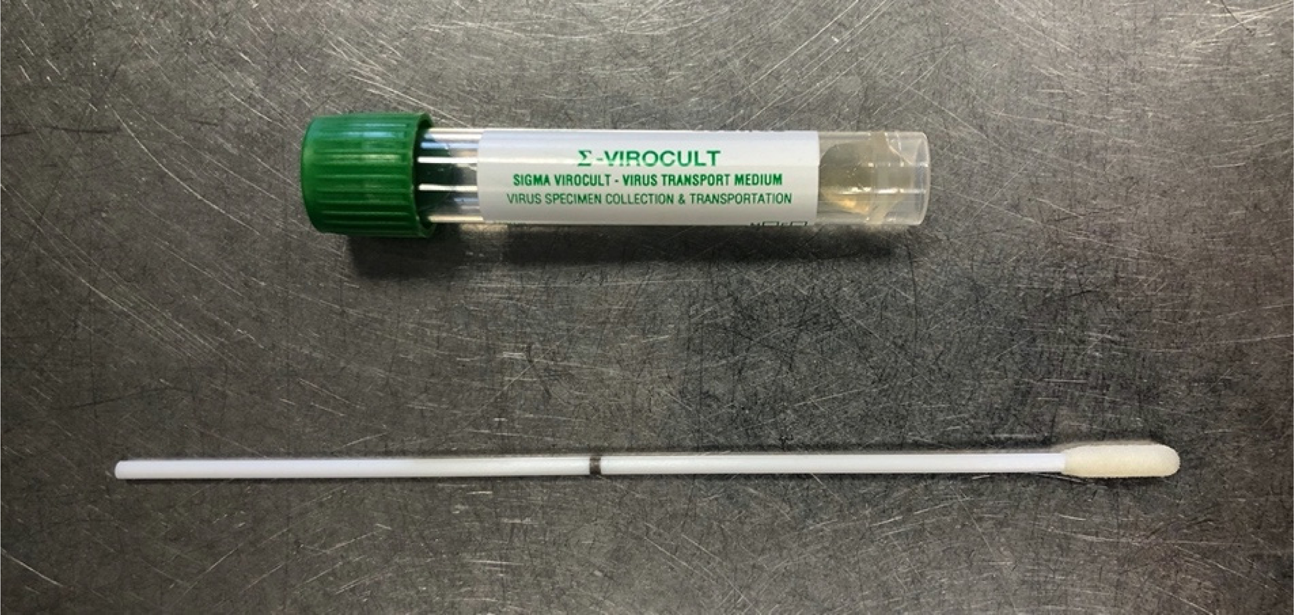
Fig. 2. Viral swab with breakpoint mechanism (black line on the swab shaft) and accompanying bottle containing virus transport medium.
The swab is then inserted into a plastic tube containing a viral transport medium (Figure 2). The manufacturer reports that this medium aids the survival of many types of common viruses, either at room temperature or refrigerated, stabilising the viral particles and utilising antimicrobial agents to prevent the growth of either bacteria or fungi that may also be present on the swab.10
The ENT-specific swab types are produced by Sigma for application in the nasopharynx.10 They utilise a finer tip and narrower shaft at the tip end of the swab. These swabs also incorporate a breakpoint feature.10
The breakpoint mechanism prevents contamination whilst transferring the shaft into the bottle; however, this feature makes the swab susceptible to fracture whilst still in the nasal cavity, as illustrated in our case. This situation could arise in non-cooperative patients, such as those with advanced dementia or severe learning difficulties, and in patients with anatomical variations (e.g. septal deviations and spurs). Head or limb movements can easily lead to the swab breaking and the swab tip being lost in the nasal cavity. If this does occur, further aerosol-generating procedures, such as flexible nasendoscopy and manual retrieval with surgical instruments, may be required in an already challenging patient.
Foreign body removal
Foreign body removal from the nose is an aerosol-generating procedure and therefore warrants robust personal protective equipment, in line with ENT UK and NHS England guidance.11,12 Covid-19 precautions also include the examination and therapeutic intervention being delivered in a ‘one-stop’ episode, limiting the location, frequency and personnel to a single episode, to reduce the risk to the patient, nearby patients and healthcare staff.11
In order to optimise the success and retrieval of a nasal foreign body in a Covid-19 patient, and reduce the risk of aerosol generation, we recommend the application of a pledget soaked in vasoconstrictor and local anaesthetic solution to the nasal cavity, to reduce mucosal oedema and desensitise nasal mucosa prior to anterior rhinoscopy. Intranasal spray should be avoided, as this may induce aerosolisation.13
Initially, anterior rhinoscopy may be carried out with a Thudicum nasal speculum, to accentuate the internal nasal valve area, the narrowest cross-sectional area of the nose. Foreign bodies deep to the internal valve require examination with a rigid or flexible nasendoscope. Close face-to-face proximity should be avoided where possible, and therefore nasal endoscopy should be carried out with a video display as per ENT UK guidance.13 Although the rigid scope is preferable, as it enables a single operator to handle both the scope and surgical instruments, it should be avoided in an uncooperative patient. A flexible nasendoscope requires two hands to operate and therefore an assistant is required for foreign body removal (Figure 3).

Fig. 3. Flexible nasendoscope and Tilley's dressing forceps in tray.
We opted to use a flexible nasendoscope with an assistant. Although this increased personnel exposure, we aimed to remove the swab at the patient's bedside in one single episode, to avoid the patient undergoing a general anaesthetic. Instrumentation to remove the foreign body is dictated by the item in question, and in this situation, we used Tilley's nasal dressing forceps to retrieve the nasal swab (Figure 3).
A retained foreign body post-procedure is a ‘never event’,14 and must be prevented at all costs. In a patient with a potential Covid-19 infection, a retained nasal swab not only poses a risk to the patient but also to healthcare staff, and the ensuing aerosol-generating procedures required to remove it further increase the risk of disease transmission to healthcare staff.
Alternative test materials
The simplest way to avoid this complication is to utilise a product without a breakpoint. Based on the information from WHO15 and other sources,Reference Druce, Garcia, Tran, Papadakis and Birch16,Reference Moore, Corden, Sinha and Jones17 there are several alternative products for viral sampling that are readily available on the market, or already in use in the clinical environment, that do not have the breakpoint feature.
The use of alternative swabs, either dry or transported with a small amount of sterile saline, may provide a useful alternative in patients at risk of experiencing premature breaking of the viral swab shaft in the nasal cavity. This is supported by the WHO guidance, which suggests that nasopharyngeal and oropharyngeal swab products with Dacron® or polyester flocked swabs can be used, and if viral transport medium is unavailable, then sterile saline may be used in replacement.15 It must be noted that when there is likely to be a delay in specimens reaching the laboratory, the use of viral transport medium is strongly recommended by WHO.15
Druce et al. demonstrated that dry swabs, transported in either saline or dry, had similar efficacy in detecting various viruses as purpose-specific viral swabs that were sent in either universal transport medium or specific viral transport medium.Reference Druce, Garcia, Tran, Papadakis and Birch16 Moore et al. revealed that the use of dry swabs with no transport medium was not only a suitable alternative, but it also produced a higher detection rate of respiratory viruses by using real-time nucleic acid sequence-based amplification, compared to more traditional methods of detection.Reference Moore, Corden, Sinha and Jones17
• Viral swab sticks generally have a breakpoint feature
• Premature activation of the breakpoint in a complex group of patients can lead to a retained swab tip in the nasal cavity
• Nasal foreign body removal is an aerosol-generating procedure and requires robust personal protective equipment
• The simplest way to avoid this complication is to utilise a product without a breakpoint
• Alternative swab products with Dacron or polyester flocked tips can be used, and if viral transport medium is unavailable, then sterile saline may be used in replacement
We suggest that hospitals liaise with their local microbiology laboratory and seek alternative arrangements for swab testing if concerned about premature breakage in vulnerable patients.
Conclusion
We have described a novel adverse event encountered with a Covid-19 viral swab. The patient was subjected to an aerosol-generating procedure to remove the nasal swab from the nasal cavity. The viral swab has an intrinsic design feature that gives rise to this complication. We have outlined appropriate techniques to address this complication and suggested a solution to circumvent this problem, which may be useful in a complex subset of suspected Covid-19 patients.
Competing interests
None declared


 Open access