COVID-19 pneumonia: The first two chest CTs in the Bamrasnaradura Infectious Disease Institute
DOI:
https://doi.org/10.46475/aseanjr.v21i2.79Keywords:
COVID-19, SARS-CoV-2, Chest computed tomography, Ground glass opacity, ConsolidationAbstract
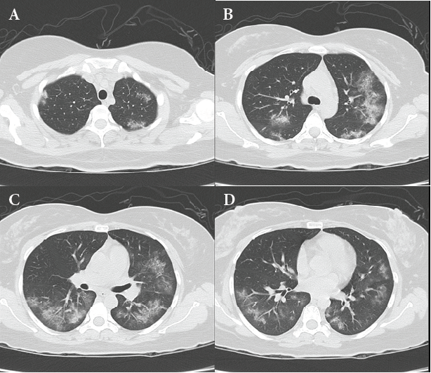
Coronavirus disease 2019 (COVID-19), caused by the severe acute respiratory syndrome coronavirus 2 (SARS-CoV-2), continues to spread rapidly around the world. We reported the first two cases of COVID-19 pneumonia who had the chest computed tomography (CT) performed at the Bamrasnaradura Infectious Disease Institute (BIDI). The chest CT findings in the two patients with COVID-19 pneumonia showed bilateral lung involvement, multifocal involvement, peripheral distribution, ground glass opacity (GGO), consolidation and GGO
with interlobular septal thickening (“crazy-paving” pattern). The chest CT findings in these patients are nonspecific and overlapped with other diseases.
Downloads
Metrics
References
World Health Organization [Internet]. Geneva: WHO; c2020 [cited 2020 Mar 20]. Naming the coronavirus disease (COVID-2019) and the virus that causes it; [about 2 screens]. Available from: https://www.who.int/emergencies/diseases/novel-coronavirus-2019/technical-guidance/naming-the-coronavirus-disease-(covid-2019)-and-the-virus-that-causes-it.
World Health Organization [Internet]. Geneva: WHO; 2020 [cited 2020 Mar 20]. Coronavirus disease 2019 (COVID-19) situation report – 51; [about 9 p.]. Available from: https://www.who.int/docs/default-source/coronaviruse/situation-reports/20200311-sitrep-51-covid-19.pdf?sfvrsn=1ba62e57_10.
Carlos WG, Dela Cruz CS, Cao B, Pasnick S, Jamil S. Novel Wuhan (2019-nCoV) coronavirus. Am J Respir Crit Care Med. 2020 Feb 15;201(4):P7-8. https://doi.org/10.1164/rccm.2014P7.
Li Q, Guan X, Wu P, Wang X, Zhou L, Tong Y, et al. Early transmission dynamics in Wuhan, China, of novel coronavirus-infected pneumonia. N Engl J Med 2020;382:1199-207. https://doi.org/10.1056/NEJMoa2001316.
Zhao S, Zhong Z, Xie X, Yu Q, Liu J. Relation between chest CT findings and clinical conditions of coronavirus disease (COVID-19) pneumonia: a multicenter study. AJR Am J Roentgenol 2020 ;214:1072-7. https://doi.org/10.2214/AJR.20.22976.
Xu X, Yu C, Qu J, Zhang L, Jiang S, Huang D, et al. Imaging and clinical features of patients with 2019 novel coronavirus SARS-CoV-2. Eur J Nucl Med Mol Imaging 2020;47:1275-80. https://doi.org/10.1007/s00259-020-04735-9.
Lai CC, Shih TP, Ko WC, Tang HJ, Hsueh PR. Severe acute respiratory syndrome coronavirus 2 (SARS-CoV-2) and coronavirus disease-2019 (COVID-19): the epidemic and the challenges. Int J Antimicrob Agents 2020 Mar;55(3):105924. https://doi.org/10.1016/j.ijantimicag.2020.105924.
Wong HYF, Lam HYS, Fong AH, Leung ST, Chin TW, Lo CSY, et al. Frequency and distribution of chest radiographic findings in COVID-19 positive patients. Radiology 2020;296(2):E72-8. https://doi.org/10.1148/radiol.2020201160.
Shi H, Han X, Jiang N, Cao Y, Alwalid O, Gu J, et al. Radiological findings from 81 patients with COVID-19 pneumonia in Wuhan, China: a descriptive study. Lancet Infect Dis 2020;20:425-34. https://doi.org/10.1016/S1473-3099(20)30086-4.
Li Y, Xia L. Coronavirus disease 2019 (COVID-19): role of chest CT in diagnosis and management. AJR Am J Roentgenol 2020;214:1280-6. https://doi.org/0.2214/AJR.20.22954.
Zhou S, Wang Y, Zhu T, Xia L. CT features of coronavirus disease 2019 (COVID-19) pneumonia in 62 patients in Wuhan, China. AJR Am J Roentgenol 2020;214:1287-94. https://doi.org/10.2214/AJR.20.22975.
Koo HJ, Lim S, Choe J, Choi SH, Sung H, Do KH. Radiographic and CT features of viral pneumonia. Radiographics 2018;38:719-39. https://doi.org/10.1148/rg.2018170048.
Franquet T. Imaging of pulmonary viral pneumonia. Radiology 2011;260:18-39. https://doi.org/10.1148/radiol.11092149.
Samir A, El-Nekiedy AAM, Baess AI, Rizk AM. H1N1 viral pneumonia: spectrum of chest HRCT findings. Egypt J Radiol Nucl Med 2016;47:1293-301.
Faria IM, Zanetti G, Barreto MM, Rodrigues RS, Araujo-Neto CA, Silva JL, et al. Organizing pneumonia: chest HRCT findings. J Bras Pneumol 2015;41:231-7.
Hosseiny M, Kooraki S, Gholamrezanezhad A, Reddy S, Myers L. Radiology perspective of coronavirus disease 2019 (COVID-19): lessons from severe acute respiratory syndrome and Middle East Respiratory Syndrome. AJR Am J Roentgenol 2020;214:1078-82. https://doi.org/10.2214/AJR.20.22969.
Ooi GC, Daqing M. SARS: radiological features. Respirology 2003;8 Suppl(Suppl 1):S15-9. https://doi.org/10.1046/j.1440-1843.2003.00519.x.
Das KM, Lee EY, Langer RD, Larsson SG. Middle East respiratory syndrome coronavirus: What does a radiologist need to know? AJR Am J Roentgenol 2016;206:1193-201. https://doi.org/10.2214/AJR.15.15363.
Pan F, Ye T, Sun P, Gui S, Liang B, Li L, et al. Time course of lung changes at chest CT during recovery from coronavirus disease 2019 (COVID-19). Radiology 2020;295:715-21. https://doi.org/10.1148/radiol.2020200370.
Memish ZA, Perlman S, Van Kerkhove MD, Zumla A. Middle East respiratory syndrome. Lancet 2020;395:1063-77. https://doi.org/10.1016/S0140-6736(19)33221-0.
Downloads
Published
How to Cite
Issue
Section
License
Copyright (c) 2020 The ASEAN Journal of Radiology

This work is licensed under a Creative Commons Attribution-NonCommercial-NoDerivatives 4.0 International License.
Disclosure Forms and Copyright Agreements
All authors listed on the manuscript must complete both the electronic copyright agreement. (in the case of acceptance)