Article Text

Statistics from Altmetric.com
Description
Several cutaneous manifestations have been seen to be associated with SARS-CoV-2 infection since the beginning of the pandemic.1–3 Acute haemorrhagic oedema of infancy (AHEI) is a vasculitis that is expressed in the first 2 years of life; it is characterised by purpuric lesions in the face, arms and legs, and is associated with oedema of the extremities. The intensity of the lesions contrasts with the benign course and good prognosis of this disease.4 5
A 14-month-old boy with a previous history of vesicoureteral reflux and recurrent pyelonephritis presented to the emergency department with a 2-day history of fever and vomiting. Lab work showed leucocytosis (21.95 × 10⁹/L), neutrophilia (11.55 × 10⁹/L), mild thrombocytopenia (172 × 109/L), elevated C reactive protein (221.8 mg/L) and normal procalcitonin (0.14 ng/mL). Urine test showed leucocyturia (237 U/L) but nitrites were negative. Therapy with cefuroxime and gentamycin was started intravenously. Although with no epidemiology or respiratory symptoms, SARS-CoV-2 was detected by reverse transcription PCR (RT-PCR) in nasopharyngeal secretions during the preadmission screening. Urine and blood cultures were negative, and chest X-ray and renal ultrasound were normal. Favourable evolution was observed, and the infant was discharged 7 days later under cefuroxime antibiotic therapy.
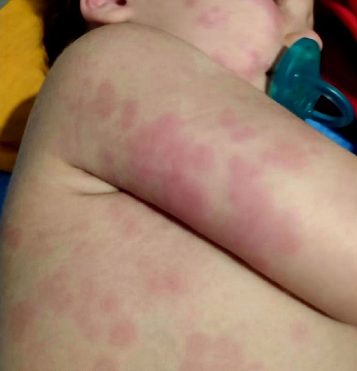
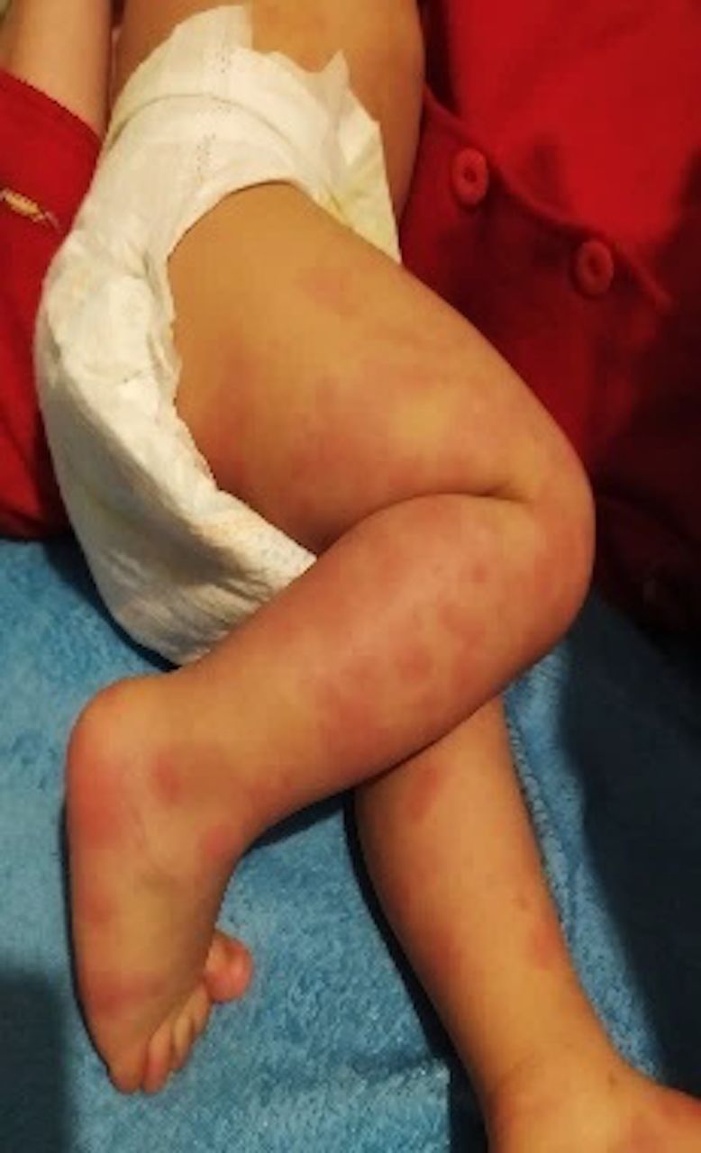
The next day, palpable maculopapular and purpuric lesions with perilesional erythema were observed, beginning in the face with progressive extension to ears, abdomen, chest (figure 1), limbs, hands, and feet; this was followed by oedema of the extremities (figure 2). Large bruise-like lesions in the arms and limbs and small purpuric lesions in the face were not blanchable regarding vitropression. The lesions were neither painful nor pruritic. No other symptoms were reported. Lab work showed leucocytes 13.34 × 10⁹/L, elevated C reactive protein 80 mg/L, sedimentation rate 74 mm/hour and D-dimers 846 ug/L. RT-PCR for PCR SARS-CoV-2 remained positive. All other respiratory viruses tested by PCR were negative. Regression of rash and oedema was observed in 3 days with full recovery after 2 weeks, only with an antihistaminic. Exclusion of allergic reaction to cefuroxime was confirmed by an oral provocation test, performed with standardised doses adjusted to the child’s weight; this test was negative.

Palpable purpuric and maculopapular lesions with perilesional erythema in acute haemorrhagic oedema of infancy (AHEI) in an infant with SARS-CoV-2 infection.

Palpable purpuric and maculopapular lesions and oedema of the extremities in acute haemorrhagic oedema of infancy (AHEI) in an infant with SARS-CoV-2 infection.
Because of the age, purpuric characteristics, swelling, localisation of the lesions (extremities, face and ears) and benign course without intern organ involvement, AHEI was the most probable diagnosis. Also, the association with prior viral infections has already been stated.5
Nevertheless, a differential diagnosis of Sweet syndrome (characterised by fever, neutrophilia and red plaques) should be considered. However, as this syndrome, usually rare in children, presents with painful tender plaques, papules and pustules, frequently associated with inflammatory disorders and malignancies,6 we ruled out this diagnose. Finally, although there were some similarities with urticarial vasculitis,7 the fact that the lesions were non-pruritic, and there were no circumscribed weals with a white centre and only residual transient hyperpigmentation, we ruled out this last diagnosis and assumed AHEI vasculitis.
This case describes the association of AHEI to SARS-CoV-2 infection as an atypical presentation of COVID-19. Therefore, this aetiology must be considered during the differential diagnosis of purpuric exanthema.
Learning points
Cutaneous lesions are an atypical manifestation of COVID-19 in children.
SARS-CoV-2 infection must be considered in the differential diagnosis of purpuric exanthema, such as acute haemorrhagic oedema of infancy.
Ethics statements
Patient consent for publication
Footnotes
Contributors BMS contributed substantially to the conception and design of the clinical case, analysed and interpreted it, and wrote the draft. MBL and ES critically revised the clinical case and included important intellectual content. AMG contributed to the design of the clinical case, took the pictures of the patient and critically revised the clinical case. All authors approved the final version submitted. BMS obtained the signature in the consent form from the caretaker of the child.
Funding The authors have not declared a specific grant for this research from any funding agency in the public, commercial or not-for-profit sectors.
Competing interests None declared.
Provenance and peer review Not commissioned; externally peer reviewed.