
COVID-19 Associated Vasculitis Confirmed by the Tissues RT-PCR: A Case Series Report
 , , ,
, , ,
Abstract
:1. Introduction
2. Materials and Methods
- -
- Epidemiology, including signs of previous COVID-19 infection (fever, cough, rhinitis, anosmia, headache, pneumonia, etc.), type of COVID-19 contact (family, class, kindergarten, or absence of evident contact), and type of COVID-19, if the corresponding assessment was performed (nasal or throat swab, PCR, IgM, or IgG against SARS-CoV-2, carried out using the enzyme-linked immunosorbent assay), in addition to the above-mentioned tissue RT-PCR;
- -
- -
- Clinical manifestations, including features of vasculitis and the following symptoms: fever, weight loss, muscle and joint pain, arthritis, rash, presence of skin or mucosal involvement, ischemia, lymphadenopathy, hepatosplenomegaly, neuropathy, and internal organ involvement (heart, lungs, brain, GI, and kidney);
- -
- Vasculitis imaging, including Doppler ultrasound, computed tomography (CT)-angiography, magnetic resonance imaging (MRI), evaluation of the caliber of involved vessels, arterial or venous thrombotic evidence, and their consequences and level of damage;
- -
- Laboratory tests, including hemoglobin, white blood cells (WBC), platelets, erythrocyte sedimentation rate (ESR), C-reactive protein (CRP), total protein, albumin, urea, creatinine, liver functional tests, ferritin, lactate dehydrogenase, coagulation status (prothrombin, fibrinogen, D-dimer), and urinalysis;
- -
- Other instrumental test results, including electrocardiogram (ECG), echocardiogram (EChoCG), abdominal ultrasound, chest CT, brain MRI, and pulmonary functional tests;
- -
- Thrombophilic genes polymorphism, which was evaluated using PCR with restriction fragment length polymorphisms according to the routine method;
- -
- Whole exome sequence to identify immunodeficiency syndromes and monogenic vasculopathies.
3. Results
3.1. COVID-19 identification
3.2. Clinical Signs of Vasculitis
3.3. Treatment
4. Discussion
5. Conclusions
Author Contributions
Funding
Institutional Review Board Statement
Informed Consent Statement
Data Availability Statement
Conflicts of Interest
Abbreviations
References
- Zhu, N.; Zhang, D.; Wang, W.; Li, X.; Yang, B.; Song, J.; Zhao, X.; Huang, B.; Shi, W.; Lu, R.; et al. A Novel Coronavirus from Patients with Pneumonia in China, 2019. N. Engl. J. Med. 2020, 382, 727–733. [Google Scholar] [CrossRef] [PubMed]
- Becker, R.C. COVID-19-associated vasculitis and vasculopathy. J. Thromb. Thrombolysis 2020, 50, 499–511. [Google Scholar] [CrossRef]
- Siddiqi, H.K.; Libby, P.; Ridker, P.M. COVID-19—A vascular disease. Trends Cardiovasc. Med. 2021, 31, 1–5. [Google Scholar] [CrossRef] [PubMed]
- Andina, D.; Belloni-Fortina, A.; Bodemer, C.; Bonifazi, E.; Chiriac, A.; Colmenero, I.; Diociaiuti, A.; El-Hachem, M.; Fertitta, L.; van Gysel, D.; et al. Skin manifestations of COVID-19 in children: Part 1. Clin. Exp. Dermatol. 2021, 46, 444–450. [Google Scholar] [CrossRef]
- Jud, P.; Gressenberger, P.; Muster, V.; Avian, A.; Meinitzer, A.; Strohmaier, H.; Sourij, H.; Raggam, R.B.; Stradner, M.H.; Demel, U.; et al. Evaluation of Endothelial Dysfunction and Inflammatory Vasculopathy after SARS-CoV-2 Infection—A Cross-Sectional Study. Front. Cardiovasc. Med. 2021, 8, 750887. [Google Scholar] [CrossRef]
- Andina, D.; Belloni-Fortina, A.; Bodemer, C.; Bonifazi, E.; Chiriac, A.; Colmenero, I.; Diociaiuti, A.; El-Hachem, M.; Fertitta, L.; van Gysel, D.; et al. Skin manifestations of COVID-19 in children: Part 3. Clin. Exp. Dermatol. 2021, 46, 462–472. [Google Scholar] [CrossRef] [PubMed]
- Henderson, L.A.; Canna, S.W.; Friedman, K.G.; Gorelik, M.; Lapidus, S.K.; Bassiri, H.; Behrens, E.M.; Ferris, A.; Kernan, K.F.; Schulert, G.S.; et al. American College of Rheumatology Clinical Guidance for Multisystem Inflammatory Syndrome in Children Associated with SARS-CoV-2 and Hyperinflammation in Pediatric COVID-19: Version 2. Arthritis Rheumatol. 2021, 73, e13–e29. [Google Scholar] [CrossRef] [PubMed]
- World Health Organization. Multisystem Inflammatory Syndrome in Children and Adolescents Temporally Related to COVID-19. 2020. Available online: https://www.who.int/news-room/commentaries/detail/multisystem-inflammatory-syndrome-in-children-and-adolescents-with-covid-19 (accessed on 15 May 2020).
- Centers for Disease Control and Prevention Health Alert Network (HAN). Multisystem Inflammatory Syndrome in Children (MIS-C) Associated with Coronavirus Disease 2019 (COVID-19). Available online: https://emergency.cdc.gov/han/2020/han00432.asp (accessed on 15 May 2020).
- You, Y.; Wang, J.; Wang, Y.; Wei, N.; Wu, L.; Chen, L.; Song, D.; Wang, Z. Non-EBV infection-associated hemophagocytic lymphohistiocytosis: A distinct subgroup where pathogen-directed therapy is essential and favorable outcomes are expected. Leuk. Lymphoma 2021, 62, 1657–1663. [Google Scholar] [CrossRef]
- Fujiwara, S.; Nakamura, H. Chronic Active Epstein-Barr Virus Infection: Is It Immunodeficiency, Malignancy, or Both? Cancers 2020, 12, 3202. [Google Scholar] [CrossRef]
- Kipkorir, V.; Cheruiyot, I.; Ngure, B.; Misiani, M.; Munguti, J. Prolonged SARS-CoV-2 RNA detection in anal/rectal swabs and stool specimens in COVID-19 patients after negative conversion in nasopharyngeal RT-PCR test. J. Med. Virol. 2020, 92, 2328–2331. [Google Scholar] [CrossRef]
- Avanzato, V.A.; Matson, M.J.; Seifert, S.N.; Pryce, R.; Williamson, B.N.; Anzick, S.L.; Barbian, K.; Judson, S.D.; Fischer, E.R.; Martens, C.; et al. Case Study: Prolonged Infectious SARS-CoV-2 Shedding from an Asymptomatic Immunocompromised Individual with Cancer. Cell 2020, 183, 1901–1912.e9. [Google Scholar] [CrossRef]
- Magro, C.M.; Mulvey, J.; Kubiak, J.; Mikhail, S.; Suster, D.; Crowson, A.N.; Laurence, J.; Nuovo, G. Severe COVID-19: A multifaceted viral vasculopathy syndrome. Ann. Diagn. Pathol. 2021, 50, 151645. [Google Scholar] [CrossRef] [PubMed]
- Colmenero, I.; Santonja, C.; Alonso-Riaño, M.; Noguera-Morel, L.; Hernández-Martín, A.; Andina, D.; Wiesner, T.; Rodríguez-Peralto, J.L.; Requena, L.; Torrelo, A. SARS-CoV-2 endothelial infection causes COVID-19 chilblains: Histopathological, immunohistochemical and ultrastructural study of seven paediatric cases. Br. J. Dermatol. 2020, 183, 729–737. [Google Scholar] [CrossRef]
- Jamiolkowski, D.; Mühleisen, B.; Müller, S.; Navarini, A.A.; Tzankov, A.; Roider, E. SARS-CoV-2 PCR testing of skin for COVID-19 diagnostics: A case report. Lancet 2020, 396, 598–599. [Google Scholar] [CrossRef] [PubMed]
- Welsh, J.D.; Hoofnagle, M.H.; Bamezai, S.; Oxendine, M.; Lim, L.; Hall, J.D.; Yang, J.; Schultz, S.; Engel, J.D.; Kume, T.; et al. Hemodynamic regulation of perivalvular endothelial gene expression prevents deep venous thrombosis. J. Clin. Investig. 2019, 129, 5489–5500. [Google Scholar] [CrossRef] [PubMed] [Green Version]
- Varatharaj, A.; Thomas, N.; Ellul, M.A.; Davies, N.W.S.; Pollak, T.A.; Tenorio, E.L.; Sultan, M.; Easton, A.; Breen, G.; Zandi, M.; et al. Neurological and neuropsychiatric complications of COVID-19 in 153 patients: A UK-wide surveillance study. Lancet Psychiatry 2020, 7, 875–882. [Google Scholar] [CrossRef] [PubMed]
- van den Berg, D.F.; Te Velde, A.A. Severe COVID-19: NLRP3 Inflammasome Dysregulated. Front. Immunol. 2020, 11, 1580. [Google Scholar] [CrossRef]
- Buckley, L.F.; Wohlford, G.F.; Ting, C.; Alahmed, A.; Van Tassell, B.W.; Abbate, A.; Devlin, J.W.; Libby, P. Role for Anti-Cytokine Therapies in Severe Coronavirus Disease 2019. Crit. Care Explor. 2020, 2, e0178. [Google Scholar] [CrossRef]
- Freeman, T.L.; Swartz, T.H. Targeting the NLRP3 Inflammasome in Severe COVID-19. Front. Immunol. 2020, 11, 1518. [Google Scholar] [CrossRef]
- Kanitakis, J.; Lesort, C.; Danset, M.; Jullien, D. Chilblain-like acral lesions during the COVID-19 pandemic (“COVID toes”): Histologic, immunofluorescence, and immunohistochemical study of 17 cases. J. Am. Acad. Dermatol. 2020, 83, 870–875. [Google Scholar] [CrossRef]
- Kolivras, A.; Dehavay, F.; Delplace, D.; Feoli, F.; Meiers, I.; Milone, L.; Olemans, C.; Sass, U.; Theunis, A.; Thompson, C.T.; et al. Coronavirus (COVID-19) infection-induced chilblains: A case report with histopathologic findings. JAAD Case Rep. 2020, 6, 489–492. [Google Scholar] [CrossRef] [PubMed]
- Batu, E.D.; Sener, S.; Ozen, S. COVID-19 associated pediatric vasculitis: A systematic review and detailed analysis of the pathogenesis. Semin. Arthritis Rheum. 2022, 55, 152047. [Google Scholar] [CrossRef] [PubMed]
- Batu, E.D.; Sener, S.; Ozomay Baykal, G.; Arslanoglu Aydin, E.; Özdel, S.; Gagro, A.; Esen, E.; Heshin-Bekenstein, M.; Akpınar Tekgöz, N.; Demirkan, F.G.; et al. The Characteristics of Patients with COVID-19-Associated Pediatric Vasculitis: An International, Multicenter Study. Arthritis Rheumatol. 2022. ahead of print. [Google Scholar] [CrossRef] [PubMed]
- Messova, A.; Pivina, L.; Muzdubayeva, Z.; Sanbayev, D.; Urazalina, Z.; Adams, A. COVID-19 and New Onset IgA Vasculitis: A Systematic Review of Case Reports. J. Emerg. Nurs. 2022, 48, 348–365. [Google Scholar] [CrossRef] [PubMed]


| ID | Sex (m/f)/ Age (y) | Previous COVID-19 (Y/N) | SARS-CoV-2 Identification (sPCR, IgM, IgG) | Type of Vasculitis | Involved Tissues | Biopsy | Systemic Inflammation/ Lab Results | Treatment | Outcomes | Ct Values for SARSCoV-2 Genes in Affected Tissues | ||||||
|---|---|---|---|---|---|---|---|---|---|---|---|---|---|---|---|---|
| ESR (mm/h) | CRP (mg/l) | Ferritin | D-dimer | ORF1 | ORF8 | N | Result | |||||||||
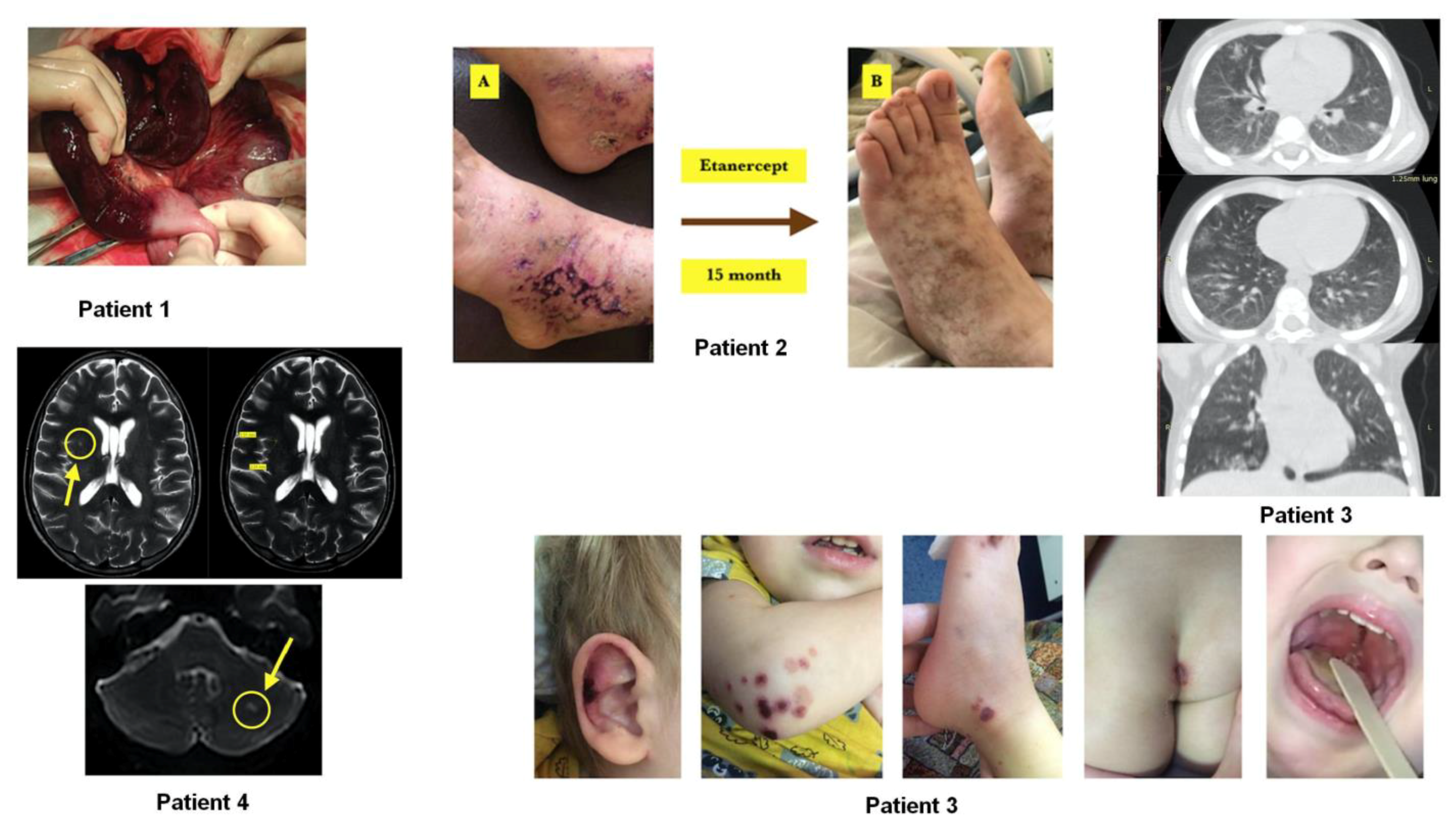
| 1 | M/16 | Y | IgG | UV | Lungs, heart, GI, TE | jejunum | 25 | 24 | 158 | 37,018 | GCS, LWH, ADA | CR | 31.6 | 30.5 | 32.0 | POS |
| 2 | F/15 | Y | sPCR, IgG | PA | Skin necrosis | skin | 15 | 4 | 17 | 237 | GCS, MTX, ETN | CR | 31.1 | 31.2 | 31.6 | POS |
| 3 | M/2 | N | IgM, IgG | IgAV/HSP | Skin necrosis, lungs, heart, GI | stomach | 16 | 23 | 27 | 2808 | GCS, CTX→MMF | PR | 32.2 | 30.5 | 32.4 | POS |
| 4 | M/15 | N | N | PA | Skin, retina, lungs, kidney, inner ear, brain, heart, GI | stomach | 23 | 153 | 432 | 1288 | GS, LWH, HCQ, TCZ, IVIG | CR | 28.6 | 27.5 | 30.1 | POS |
Disclaimer/Publisher’s Note: The statements, opinions and data contained in all publications are solely those of the individual author(s) and contributor(s) and not of MDPI and/or the editor(s). MDPI and/or the editor(s) disclaim responsibility for any injury to people or property resulting from any ideas, methods, instructions or products referred to in the content. |
© 2023 by the authors. Licensee MDPI, Basel, Switzerland. This article is an open access article distributed under the terms and conditions of the Creative Commons Attribution (CC BY) license (https://creativecommons.org/licenses/by/4.0/).
Share and Cite
Belozerov, K.E.; Avrusin, I.S.; Andaryanova, L.I.; Guseva, A.M.; Shogenova, Z.S.; Belanovich, I.N.; Lobacheva, A.V.; Kornishina, T.L.; Isupova, E.A.; Masalova, V.V.; et al. COVID-19 Associated Vasculitis Confirmed by the Tissues RT-PCR: A Case Series Report. Biomedicines 2023, 11, 870. https://doi.org/10.3390/biomedicines11030870
Belozerov KE, Avrusin IS, Andaryanova LI, Guseva AM, Shogenova ZS, Belanovich IN, Lobacheva AV, Kornishina TL, Isupova EA, Masalova VV, et al. COVID-19 Associated Vasculitis Confirmed by the Tissues RT-PCR: A Case Series Report. Biomedicines. 2023; 11(3):870. https://doi.org/10.3390/biomedicines11030870
Chicago/Turabian StyleBelozerov, Konstantin E., Ilia S. Avrusin, Lyubov I. Andaryanova, Anna M. Guseva, Zaira S. Shogenova, Irina N. Belanovich, Anna V. Lobacheva, Tatiana L. Kornishina, Eugenia A. Isupova, Vera V. Masalova, and et al. 2023. "COVID-19 Associated Vasculitis Confirmed by the Tissues RT-PCR: A Case Series Report" Biomedicines 11, no. 3: 870. https://doi.org/10.3390/biomedicines11030870