
An Atypical Case of Aphasia: Transitory Ischemic Attack in a 13-Year-Old Patient with Asymptomatic SARS-CoV-2 Infection

 , , , , , and
Abstract
:1. Introduction
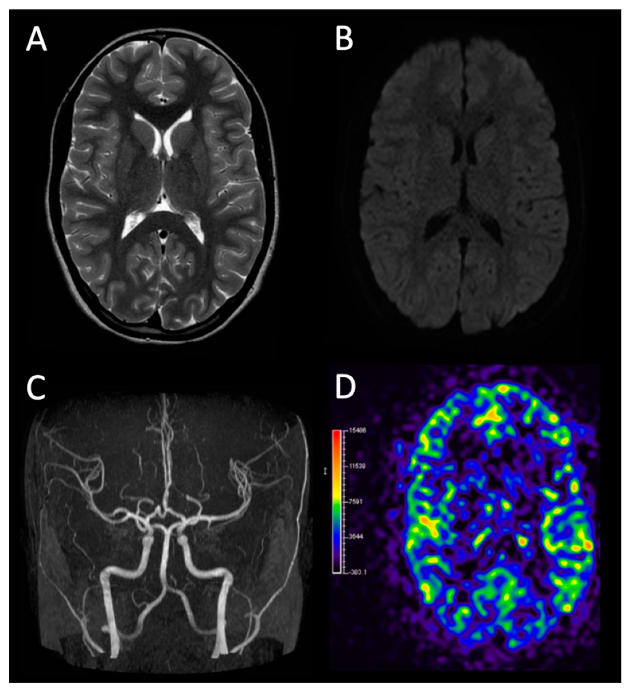
2. Case
3. Differential Diagnosis and Discussion
3.1. Transient Ischemic Attack from Patent Oval Foramen
3.2. Transient Ischemic Attack of Infectious Etiology
4. Conclusions and Take-Home Message
Author Contributions
Funding
Institutional Review Board Statement
Informed Consent Statement
Acknowledgments
Conflicts of Interest
References
- Quintanilla-Sánchez, C.; Salcido-Montenegro, A. Acute cerebrovascular events in severe and nonsevere COVID-19 patients: A systematic review and meta-analysis. Rev. Neurosci. 2022. [Google Scholar] [CrossRef] [PubMed]
- Nalleballe, K.; Reddy Onteddu, S. Spectrum of neuropsychiatric manifestations in COVID-19. Brain Behav. Immunity 2020, 88, 71–74. [Google Scholar] [CrossRef] [PubMed]
- Garg, D.; Srivastava, A.K. Beyond Fever, Cough and Dyspnea: The Neurology of COVID-19. J. Assoc. Physicians India 2020, 68, 62–66. [Google Scholar] [PubMed]
- Karimi, L.; Sales, C. Acute Ischemic Stroke in SARS-CoV, MERS-CoV, SARS-CoV-2: Neurorehabilitation Implications of Inflammation Induced Immunological Responses Affecting Vascular Systems. Front. Neurol. 2020, 11, 565665. [Google Scholar] [CrossRef]
- Finsterer, J.; Wilfing, A. Anticoagulated de novo atrial flutter complicated by transitory ischemic attack in fatal COVID-19. Clin. Case Rep. 2022, 10, e05246. [Google Scholar] [CrossRef]
- Choudhry, H.; Klingensmith, J.; Border, D.L.; Myers, M.; Mercado, E. Large Middle Cerebral Artery Ischemic Stroke in a Therapeutically Anticoagulated Patient with Severe SARS-CoV-2 Infection. Neurologist 2021. [Google Scholar] [CrossRef]
- Schober, M.E.; Robertson, C.L. COVID-19 and the Pediatric Nervous System: Global Collaboration to Meet a Global Need. Neurocrit. Care 2021, 35, 283–290. [Google Scholar] [CrossRef]
- LaRovere, K.L.; Riggs, B.J. Neurologic involvement in children and adolescents hospitalized in the United States for COVID-19 or multisystem infammatory syndrome. JAMA Neurol. 2021, 78, 536–547. [Google Scholar] [CrossRef]
- Ranabothu, S.; Onteddu, S. Spectrum of COVID-19 in children. Acta Paediatr. 2020, 109, 1899–1900. [Google Scholar] [CrossRef]
- Panda, P.K.; Sharawat, I.K. Neurological complications of SARS-CoV-2 infection in children: A systematic review and meta-analysis. J. Trop. Pediatr. 2020, 67, fmaa070. [Google Scholar] [CrossRef]
- Dalakas, M.C. Guillain-Barre syndrome: The first documented COVID19-triggered autoimmune neurologic disease: More to come with myositis in the ofng. Neurol. Neuroimmunol. Neuroinfamm. 2020, 7, e781. [Google Scholar] [CrossRef] [PubMed]
- Ludvigsson, J.F. Systematic review of COVID-19 in children shows milder cases and a better prognosis than adults. Acta Paediatr. 2020, 109, 1088–1095. [Google Scholar] [CrossRef] [PubMed]
- Hosey, M.M.; Needham, D.M. Survivorship after COVID-19 ICU stay. Nat. Rev. Dis. Primers 2020, 6, 60. [Google Scholar] [CrossRef]
- Garrigues, E.; Janvier, P. Post-discharge persistent symptoms and health-related quality of life after hospitalization for COVID-19. J. Infect. 2020, 81, e4–e6. [Google Scholar] [CrossRef] [PubMed]
- Almeria, M.; Cejudo, J.C. Cognitive profile following COVID-19 infection: Clinical predictors leading to neuropsychological impairment. Brain Behav. Immun. Health 2020, 9, 100163. [Google Scholar] [CrossRef] [PubMed]
- Yew, K.S.; Cheng, E.M. Diagnosis of Acute Stroke. Am. Fam. Physician 2015, 91, 528–536. [Google Scholar]
- Miranda, B.; Fonseca, A.C. Patent foramen ovale and stroke. J. Neurol. 2018, 265, 1943–1949. [Google Scholar] [CrossRef]
- García-Azorín, D.; Abenza Abildúa, M.J. Neurological presentations of COVID-19: Findings from the Spanish Society of Neurology neuroCOVID-19 registry. Spanish neuroCOVID registry group. J. Neurol. Sci. 2021, 423, 117283. [Google Scholar] [CrossRef]
- Koh, J.S.; De Silva, D.A. Neurology of COVID-19 in Singapore. J. Neurol. Sci. 2020, 418, 117118. [Google Scholar] [CrossRef]
- Mantero, V.; Basilico, P. Recurrent Transient Ischemic Attack in a Young Patient with COVID-19. J. Clin. Neurol. 2020, 16, 513–514. [Google Scholar] [CrossRef]
- Singer, T.G.; Evankovich, K.D. Coronavirus Infections in the Nervous System of Children: A Scoping Review Making the Case for Long-Term Neurodevelopmental Surveillance. Pediatr. Neurol. 2021, 117, 47–63. [Google Scholar] [CrossRef] [PubMed]
- Ray, S.T.J.; Abdel-Mannan, O. Neurological manifestations of SARS-CoV-2 infection in hospitalised children and adolescents in the UK: A prospective national cohort study. Lancet Child Adolesc. Health 2021, 5, 631–641. [Google Scholar] [CrossRef]
- Iadecola, C.; Anrather, J. Efects of COVID-19 on the nervous system. Cell 2020, 183, 16–27. [Google Scholar] [CrossRef] [PubMed]
- Anoop, U.R.; Verma, K. Happy hypoxemia in COVID-19-a neural hypothesis. ACS Chem. Neurosci. 2020, 11, 1865–1867. [Google Scholar]
- Lukiw, W.J.; Pogue, A.; Hill, J.M. SARS-CoV-2 Infectivity and Neurological Targets in the Brain. Cell. Mol. Neurobiol. 2022, 42, 217–224. [Google Scholar] [CrossRef]
- Yang, S.; Jin, H. Diverse functions and mechanisms of pericytes in ischemic stroke. Curr. Neuropharmacol. 2017, 15, 892–905. [Google Scholar] [CrossRef] [Green Version]
- Wild, J.R.L.; Staton, C.A. Neuropilins: Expression and roles in the epithelium. Int. J. Exp. Pathol. 2012, 93, 81–103. [Google Scholar] [CrossRef]
- Robinson, F.A.; Mihealsick, R.P. Role of angiotensin converting enzyme 2 and pericytes in cardiac complications of COVID-19 infection. Am. J. Physiol. Heart Circ. Physiol. 2020, 319, H1059–H1068. [Google Scholar] [CrossRef]
- Abdel-Mannan, O.; Eyre, M. Neurologic and radiographic findings associated with COVID-19 infection in children. JAMA Neurol. 2020, 77, 1440–1445. [Google Scholar] [CrossRef]
- Schurink, B.; Roos, E. Viral presence and immunopathology in patients with lethal COVID-19: A prospective autopsy cohort study. Lancet Microbe 2020, 1, e290–e299. [Google Scholar] [CrossRef]
- Das, S.; Ray, B.K. Impact of COVID-19 pandemic in natural course of Moyamoya Angiopathy: An experience from tertiary-care-center in India. Egypt J. Neurol. Psychiatr. Neurosurg. 2021, 57, 166. [Google Scholar] [CrossRef] [PubMed]


| Blood Tests | |||
|---|---|---|---|
| Units of Measurement | Normal Reference Range | Result | |
| AUTOANTIBODIES (Ab) | |||
| Anti Cardiolipin Ab | 0–0 | 5.3 | |
| Anti beta2-Glicoprotein 1 Ab | 0–0 | 2 | |
| Anti Transglutaminase Ab | UR/mL | 0–19 | <2 |
| SERUM EXAMINATION | |||
| Sodium | mEq/L | 135–145 | 138 |
| Potassium | mEq/L | 3.5–5.1 | 4.3 |
| Calcium | mEq/L | 4.05–5.2 | 4.75 |
| Chlorine | mEq/L | 98–110 | 105 |
| Bicarbonates | mEq/L | 20–25 | 21.9 |
| Anionic gap | 7–14 | 11 | |
| Azotemia | mg/dL | 15–40 | 27 |
| Blood glucose | mg/dL | 60–100 | 88 |
| Magnesium | mg/dL | 1.7–2.55 | 2.05 |
| Phosphorus | mg/dL | 3–5.6 | 3.84 |
| Creatinin | mg/dL | 0.4–1.2 | 0.72 |
| AST transaminase | U/L | 0–35 | 16 |
| ALT transaminase | U/L | 0–35 | 8 |
| Gamma-glutamyl transferase (GGT) | U/L | 8–32 | 8 |
| Alkaline phosphatase | UI/L | 57–254 | 110 |
| Conjugated bilirubin | mg/dL | 0–0.3 | 0.61 |
| Unconjugated bilirubin | mg/dL | 0–99.99 | 1.79 |
| Total bilirubin | mg/dL | 0–1 | 2.40 |
| Triglycerides | mg/dL | 30–160 | 70 |
| Total cholesterol | mg/dL | 80–180 | 128 |
| HDL cholesterol | mg/dL | 65–100 | 69 |
| LDL cholesterol | mg/dL | <100 | 59 |
| Homocysteine | umol/L | 0–10 | 8.5 |
| Beta2-microglobulin | mg/L | 0–2.2 | 1.9 |
| Hemolysis index | 0–0 | absent | |
| Jaundice index | 0–0 | absent | |
| Lipemic index | 0–0 | absent | |
| COAGULATION STUDY | |||
| Prothrombin activity | % | 63–129 | 100.7 |
| Prothrombin activity | 0.74–1.25 | 0.990 | |
| Partial thromboplastin ratio | 0.86–1.23 | 1.03 | |
| Partial thromboplastin time | s. | 23.2–33.2 | 27.8 |
| Fibrinogen | mg/100mL | 180–350 | 238 |
| Antithrombin | % | 75–125 | 109 |
| D-dimer | mg/L FEU | 0–0.55 | <0.55 |
| C protein | % | 70–140 | 103 |
| LAC | 0–1.3 | 1.20 | |
| Factor VIII activity | % | 50–150 | 124.75 |
| VON WILLEBRAND factor activity (Ristocetinal cofactor) | % | 46–173 | 68 |
| Mutated FII | Absent | ||
| Leiden FV | Absent | ||
| VON WILLEBRAND factor antigen | % | 50–160 | 62.36 |
| BLOOD COUNT | |||
| Hematocrit | % | 38–46 | 40.4 |
| Hemoglobin | g/dL | 11.5–16.5 | 14.2 |
| Erythroblasts | % | 0–0 | 0.0 |
| Erythroblasts | ×103/uL | 0–0 | 0.0 |
| Red blood cells | ×106/uL | 3.9–5.6 | 4.52 |
| White blood cells | ×103/uL | 4–9.8 | 8.06 |
| MCHC | g/dL | 32–36 | 35.2 |
| MCH | pg | 27–32 | 31.4 |
| MCV | fL | 82–96 | 89.3 |
| MPV | fL | 7–12 | 9.7 |
| Platelets | ×103/uL | 150–450 | 167 |
| RDW | % | 11.5–16 | 13.0 |
| Basophils | % | 0–1 | 0.3 |
| Basophils | ×103/uL | 0–0.08 | 0.02 |
| Eosinophils | ×103/uL | 0–0.62 | 0.22 |
| Eosinophils | % | 0–9.3 | 2.7 |
| Lymphocytes | ×103/uL | 1.37–6.81 | 1.69 |
| Lymphocytes | % | 20.7–50.4 | 21.0 |
| Large unstained cells (LUC) | ×103/uL | 0–0.24 | 0.12 |
| Large unstained cells (LUC) | % | 0–3.4 | 1.5 |
| Monocytes | ×103/uL | 0.24–0.71 | 0.39 |
| Monocytes | % | 3.9–9 | 4.9 |
| Neutrophils | ×103/uL | 2.1–6.43 | 5.61 |
| Neutrophils | % | 35.5–70.8 | 69.6 |
| INFLAMMATION INDICES | |||
| C reactive protein | mg/dL | 0–0.46 | <0.46 |
| HORMONAL TESTS | |||
| FT4 | pg/mL | 9.3–17 | 11.4 |
| TSH | uU/mL | 0.2–4.2 | 1.660 |
| IMMUNITY STUDY | |||
| Serum IgA | mg/dL | 70–400 | 38 |
| Serum IgG | mg/dL | 700–1600 | 1115 |
| Serum IgM | mg/dL | 40–230 | 69.0 |
| Serum IgM anti-2019-nCoV | AU/mL | 0–1 | <1 |
| Serum IgG anti-2019-nCoV | AU/mL | <1.1 | 18.0 |
| Blood Exams | Instrumental Examinations |
|---|---|
| Complete blood count and leukocyte count | Brain MRI |
| Complete serum examination with inflammatory indices | Electroencephalogram |
| Indices of thyroid function and celiac disease screening | Electrocardiogram and echocardiogram |
| Extended study of coagulation | Ecocolordoppler exam of lower limbs, abdomen vessels and supra-aortic trunks |
| Anti-phospholipid autoantibodies | Echocardiogram with bubble study |
Publisher’s Note: MDPI stays neutral with regard to jurisdictional claims in published maps and institutional affiliations. |
© 2022 by the authors. Licensee MDPI, Basel, Switzerland. This article is an open access article distributed under the terms and conditions of the Creative Commons Attribution (CC BY) license (https://creativecommons.org/licenses/by/4.0/).
Share and Cite
Scaglione, M.; Napoli, F.; Prato, G.; Severino, M.; Bertamino, M.; Signa, S.; Maghnie, M. An Atypical Case of Aphasia: Transitory Ischemic Attack in a 13-Year-Old Patient with Asymptomatic SARS-CoV-2 Infection. Children 2022, 9, 983. https://doi.org/10.3390/children9070983
Scaglione M, Napoli F, Prato G, Severino M, Bertamino M, Signa S, Maghnie M. An Atypical Case of Aphasia: Transitory Ischemic Attack in a 13-Year-Old Patient with Asymptomatic SARS-CoV-2 Infection. Children. 2022; 9(7):983. https://doi.org/10.3390/children9070983
Chicago/Turabian StyleScaglione, Marco, Flavia Napoli, Giulia Prato, Mariasavina Severino, Marta Bertamino, Sara Signa, and Mohamad Maghnie. 2022. "An Atypical Case of Aphasia: Transitory Ischemic Attack in a 13-Year-Old Patient with Asymptomatic SARS-CoV-2 Infection" Children 9, no. 7: 983. https://doi.org/10.3390/children9070983