 Daiana Mir1†
 Natalia Rego2†
 Paola Cristina Resende3†Fernando Tort4†Tamara Fernández-Calero2,5†
 Verónica Noya2,6Mariana Brandes2Tania Possi6Mailen Arleo6Natalia Reyes6Matías Victoria4Andres Lizasoain4
 Matías Castells4Leticia Maya4Matías Salvo4
 Tatiana Schäffer Gregianini7Marilda Tereza Mar da Rosa7Letícia Garay Martins8
 Cecilia Alonso9Yasser Vega10Cecilia Salazar11Ignacio Ferrés11
 Pablo Smircich12Jose Sotelo Silveira13
 Rafael Sebastián Fort12
 Cecilia Mathó13
 Ighor Arantes14
 Luciana Appolinario3
 Ana Carolina Mendonça3
 María José Benítez-Galeano1Camila Simoes2
 Martín Graña2
 Fernando Motta3
 Marilda Mendonça Siqueira3
 Gonzalo Bello14*
 Rodney Colina4*
 Lucía Spangenberg2,15*
- 1Unidad de Genómica y Bioinformática, Departamento de Ciencias Biológicas, Centro Universitario Regional Litoral Norte, Universidad de la República, Salto, Uruguay
- 2Unidad de Bioinformaítica, Institut Pasteur de Montevideo, Montevideo, Uruguay
- 3Laboratório de Vírus Respiratórios e Sarampo, Instituto Oswaldo Cruz – Fiocruz, Rio de Janeiro, Brazil
- 4Laboratorio de Virología Molecular, Departamento de Ciencias Biológicas, Centro Universitario Regional Litoral Norte, Universidad de la República, Salto, Uruguay
- 5Departamento de Ciencias Exactas y Naturales, Universidad Católica del Uruguay, Montevideo, Uruguay
- 6Laboratorio de Biología Molecular, Sanatorio Americano, Montevideo, Uruguay
- 7Laboratório Central de Saúde Pública, Centro Estadual de Vigilância em Saúde da Secretaria de Saúde do Estado do Rio Grande do Sul (LACEN/CEVS/SES-RS), Porto Alegre, Brazil
- 8Centro Estadual de Vigilância em Saúde da Secretaria de Saúde do Estado do Rio Grande do Sul, Porto Alegre, Brazil
- 9CENUR Este-Sede Rocha-Universidad de la República, Montevideo, Uruguay
- 10Laboratorio DILAVE/MGAP-INIA-Universidad de la República, Tacuarembó, Uruguay
- 11Laboratorio de Genómica Microbiana, Institut Pasteur de Montevideo, Montevideo, Uruguay
- 12Departamento de Genómica, Instituto de Investigaciones Biológicas Clemente Estable, MEC, Laboratorio de Interacciones Moleculares, Facultad de Ciencias, Universidad de la República, Montevideo, Uruguay
- 13Departamento de Genómica, Instituto de Investigaciones Biológicas Clemente Estable, MEC. Sección Biología Celular, Departamento de Biología Celular y Molecular, Facultad de Ciencias, Universidad de la República, Montevideo, Uruguay
- 14Laboratorio de AIDS e Imunologia Molecular, Instituto Oswaldo Cruz – Fiocruz, Rio de Janeiro, Brazil
- 15Departamento de Informática y Ciencias de la Computación, Universidad Católica del Uruguay, Montevideo, Uruguay
Uruguay is one of the few countries in the Americas that successfully contained the coronavirus disease 19 (COVID-19) epidemic during the first half of 2020. Nevertheless, the intensive human mobility across the dry border with Brazil is a major challenge for public health authorities. We aimed to investigate the origin of severe acute respiratory syndrome coronavirus 2 (SARS-CoV-2) strains detected in Uruguayan localities bordering Brazil as well as to measure the viral flux across this ∼1,100 km uninterrupted dry frontier. Using complete SARS-CoV-2 genomes from the Uruguayan–Brazilian bordering region and phylogeographic analyses, we inferred the virus dissemination frequency between Brazil and Uruguay and characterized local outbreak dynamics during the first months (May–July) of the pandemic. Phylogenetic analyses revealed multiple introductions of SARS-CoV-2 Brazilian lineages B.1.1.28 and B.1.1.33 into Uruguayan localities at the bordering region. The most probable sources of viral strains introduced to Uruguay were the Southeast Brazilian region and the state of Rio Grande do Sul. Some of the viral strains introduced in Uruguayan border localities between early May and mid-July were able to locally spread and originated the first outbreaks detected outside the metropolitan region. The viral lineages responsible for Uruguayan urban outbreaks were defined by a set of between four and 11 mutations (synonymous and non-synonymous) with respect to the ancestral B.1.1.28 and B.1.1.33 viruses that arose in Brazil, supporting the notion of a rapid genetic differentiation between SARS-CoV-2 subpopulations spreading in South America. Although Uruguayan borders have remained essentially closed to non-Uruguayan citizens, the inevitable flow of people across the dry border with Brazil allowed the repeated entry of the virus into Uruguay and the subsequent emergence of local outbreaks in Uruguayan border localities. Implementation of coordinated bi-national surveillance systems is crucial to achieve an efficient control of the SARS-CoV-2 spread across this kind of highly permeable borderland regions around the world.
Introduction
The severe acute respiratory syndrome coronavirus 2 (SARS-CoV-2), the causative agent of coronavirus disease 19 (COVID-19), was first reported in South America on February 26, 2020 and rapidly spread through the region. South America is actually the second-worst affected region in the world, with more than 11 million SARS-CoV-2 cases and nearly 350,000 deaths confirmed as of December, 2020 (Worldometers, 2020). While the virus exponentially spread during the first half of 2020 in most South American countries, the rapid implementation of non-pharmaceutical interventions avoided an exponential growth of SARS-CoV-2 cases in Uruguay (Moreno et al., 2020; Valcarcel et al., 2020). Six months after the first four cases were reported on March 13, 2020, the country registered the lowest total and per capita numbers of SARS-CoV-2 cases (1,808 cases, 520 cases/million inhabitants) and deaths (45 deaths, 13 deaths/million inhabitants) in the region (Worldometers, 2020).
Despite Uruguay’s success to control the early expansion of SARS-CoV-2, the intensive cross-border human mobility between Uruguay and neighboring countries heavily affected by the pandemic is a major concern for public health authorities, aiming to achieve long-term epidemic control. With an area of approximately 176,000 km2 and 3.5 million inhabitants, Uruguay borders with Argentina to its west and southwest and with Brazil to its north and east. Of particular concern is the border with Brazil, a porous ∼1,100 km strip of land that separates the southernmost Brazilian state of Rio Grande do Sul (RS) and the Uruguayan departments of Artigas (AR), Rivera (RI), Cerro Largo (CL), Treinta y Tres (TT), and Rocha (RO). The Brazilian–Uruguayan border hosts about 170,000 people that live in twin cities located both sides of a dry border and that maintain an intense economic and social interdependence (Arnson et al., 2020).
With a total area of about 282,000 km2 and 11.3 million inhabitants, RS is the fifth-most-populous Brazilian state and as of July 31, 2020, registered 66,692 SARS-CoV-2 cases and 1,876 deaths (saude, 2021). After an initial phase of relatively slow growth, the COVID-19 epidemic displayed a sharp increase in RS since early May that coincides with the detection of several outbreaks along Uruguayan border departments. The largest SARS-CoV-2 Uruguayan outbreaks (∼50–100 confirmed cases), outside the metropolitan region during the first months after the first detected cases in the country, were at RI and TT departments in May and June, respectively, while smaller outbreaks (∼10–20 confirmed cases) were detected in AR and CL by July (github, 2020). As of July 31, 2020, a total of 201 SARS-CoV-2 cases were reported in Uruguayan municipalities located along the border with Brazil, which represents 16% of the laboratory-confirmed cases in the country (github, 2020).
Previous phylogenetic analyses revealed the circulation of different predominant SARS-CoV-2 lineages in Brazil (B.1.1.28 and B.1.1.33) (Candido et al., 2020; Resende et al., 2020b) and Uruguay (A.5 and B.1) (Elizondo et al., 2020; Salazar et al., 2020), thus supporting independent viral seeding events and little viral exchanges between these neighboring countries during the very early phase of the epidemic. However, most Uruguayan SARS-CoV-2 samples previously analyzed were from Montevideo, the capital city of the country, and there is only scarce information concerning the virus strains circulating in Uruguayan localities bordering Brazil.
We generated 122 SARS-CoV-2 whole-genome sequences recovered from cases isolated at Uruguayan border departments (n = 54) as well as in the southernmost Brazilian state of RS (n = 68) to gain insight into the origin and dynamics of SARS-CoV-2 spread at the Brazilian–Uruguayan border. Our study provides important findings that demonstrate the relevance of building bi-national genomic surveillance workforces in countries with porous and dynamic bordering regions.
Materials and Methods
SARS-CoV-2 Samples and Ethical Aspects
A total of 122 SARS-CoV-2 whole-genomes were recovered from nasopharyngeal–throat combined swab samples collected from deceased cases (n = 5), clinically ill or asymptomatic individuals that reside in five different Uruguayan departments (n = 54) at the Brazilian border and 41 different municipalities of the RS Brazilian state (n = 68) (Figure 1 and Supplementary Table 1 in Supplementary Material Section 2). Uruguayan samples were collected between May 5 and July 26, 2020, and underwent testing at the Universidad de la República, CENUR Litoral Norte, Salto (Molecular Virology Lab); Sanatorio Americano Montevideo (SASA, Molecular Biology Lab); Universidad de la República CURE Este, Rocha (Molecular Ecology Lab); and Laboratory of DILAVE/MGAP-INIA-UdelaR (Tacuarembó). Sequencing of the Uruguayan samples was held at the Institut Pasteur de Montevideo (IPMON, Bioinformatics Unit) and Instituto de Investigaciones Biológicas Clemente Estable (IIBCE, Genomics Department). Brazilian samples were recovered from sentinel locations in RS state from March 9 to August 16, 2020, and sent to the central laboratory from RS state (LACEN-RS) for SARS-CoV-2 RT-PCR detection. Positive Brazilian samples were subsequently sent to the Laboratory of Respiratory Viruses and Measles, IOC, FIOCRUZ, WHO Regional Reference Laboratory for Coronavirus in the Americas. This study was approved by Ethics Committees in Uruguay (SASA Ethics Committee: CEI-SASA; Supplementary Material Section 4) and Brazil (FIOCRUZ-IOC Ethics Committee: 68118417.6.0000.5248 and Brazilian Ministry of Health SISGEN: A1767C3).
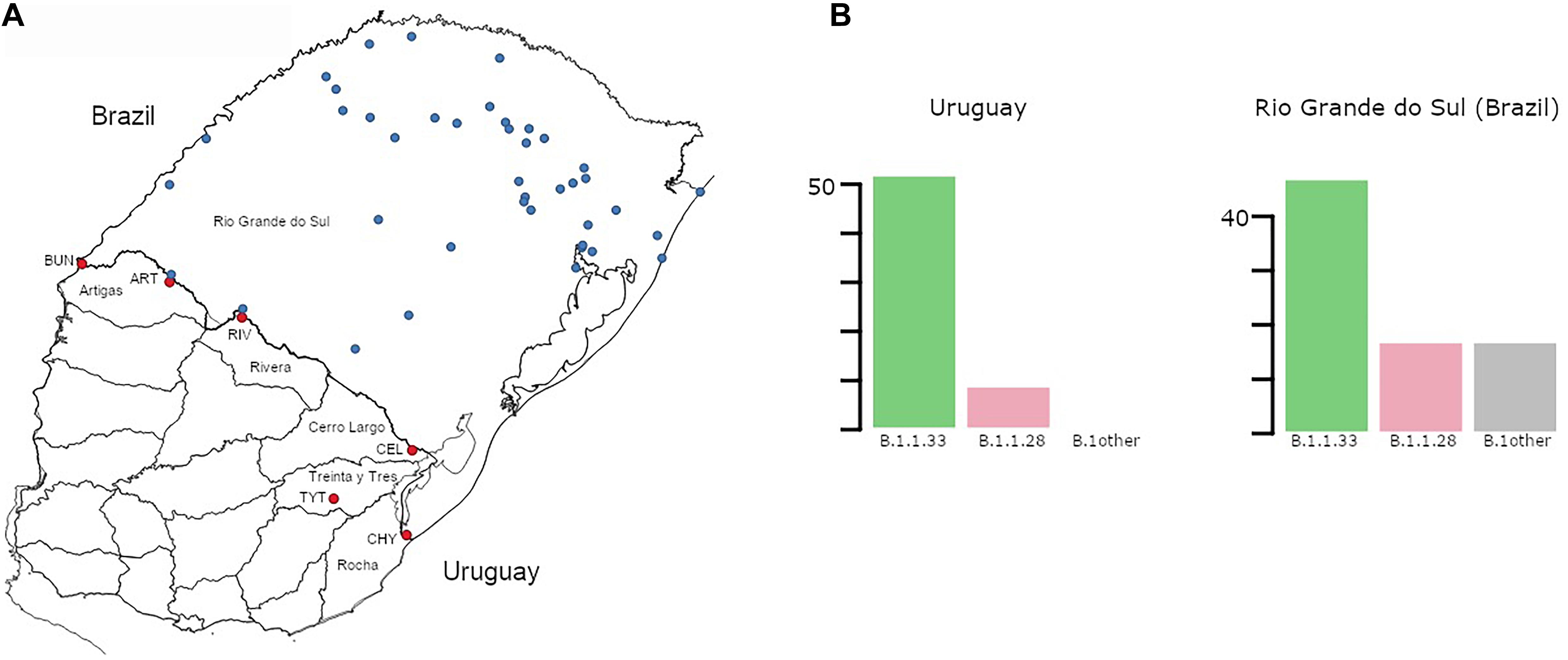
Figure 1. (A) Map of Uruguay and Rio Grande do Sul (Brazil) showing the distribution of samples included in the study. Sampling locations in Uruguay and Rio Grande do Sul (RS) are marked in red and blue, respectively. Uruguayan departments bordering Brazil are explicitly named. Additionally, towns/cities within those departments are marked: BUN, Bella Unión; ART, Artigas; RIV, Rivera; CEL, Rio Branco; TYT, Treinta y Tres; CHY, Chuy. (B) Prevalence (number of cases) of the different SARS-CoV-2 lineages detected in Uruguayan departments at the border region and in RS. While no “B.1other” were found in Uruguay, 6 B.1 and 7 B.1.1 samples were found in RS. The total number of cases for Uruguay was 59 and for RS 81.
SARS-CoV-2 Identification of Positive Samples, Amplification, and Sequencing
Molecular detection of the virus was performed with different kits at each center (Bio-Manguinhos SARS-CoV-2 kit, OneStep RT-qPCR kit COVID-19 RT-PCR Real TM Fast, Coronavirus COVID-19 genesig Real-Time PCR assay), according to manufacturer’s instructions (Supplementary Material Section 1). SARS-CoV-2 genomes were recovered using both long and short PCR amplicon protocols (Quick, 2020; Resende, 2020; Resende et al., 2020a) and three sequencing technologies: Oxford Nanopore Technologies (ONT), Illumina, and Ion Torrent (Supplementary Table 1 in Supplementary Material Section 2). See Supplementary Material Section 1 for details.
SARS-CoV-2 Whole-Genome Consensus Sequences
Whole-genome consensus sequences obtained from ONT were generated using an adaptation of the nCoV-2019 novel coronavirus ARTIC bioinformatics protocol1 as in Resende et al. (2020c) with adjustments that are available at https://github.com/iferres/ncov2019-artic-nf. For Illumina and Ion Torrent, BCFtools was used for single-nucleotide polymorphism (SNP) calling (mpileup), SNP filtering, and consensus sequences reconstruction (consensus function) as described in detail in the Supplementary Material Section 1. Positions of interest were manually inspected to resolve undetermined bases. All genomes obtained in this study were uploaded at the EpiCoV database in the GISAID initiative under the accession numbers EPI_ISL_729794 to EPI_ISL_729861 (Brazilian genomes), and accession numbers of Uruguayan genomes are in Supplementary Table 2 in Supplementary Material Section 2.
SARS-CoV-2 Genotyping and Maximum Likelihood Phylogenetic Analyses
Uruguayan and Brazilian SARS-CoV-2 genome sequences were initially assigned to viral lineages according to Rambaut et al. (2020), using the pangolin web application2 and later confirmed using maximum likelihood (ML) phylogenetic analyses. ML phylogenetic analyses were performed with the PhyML 3.0 program (Guindon et al., 2010), using an online web server (Guindon et al., 2005; Supplementary Material Section 1). Branch support was assessed by the approximate likelihood-ratio test based on a Shimodaira–Hasegawa-like procedure (SH-aLRT) with 1,000 replicates (Anisimova and Gascuel, 2006).
Phylogeographic Analyses
The previously generated ML trees were employed for the ancestral character state reconstruction (ACR) of epidemic locations with PastML (Ishikawa et al., 2019), using the marginal posterior probabilities approximation (MPPA) method with an F81-like model. We next constructed a time-scaled Bayesian phylogenetic tree for the Brazilian and Uruguayan sequences belonging to the B.1.1.28 and B.1.1.33 lineages using the Bayesian Markov chain Monte Carlo (MCMC) approach implemented in BEAST 1.10 (Suchard et al., 2018) with BEAGLE library v3 to improve computational time (Supplementary Material Section 1). Viral migrations were reconstructed using a reversible discrete phylogeographic model with a continuous-time Markov chain (CTMC) rate reference prior (Ferreira and Suchard, 2008).
Within-Host Diversity
Only those synapomorphic sites with at least 100 reads were kept for further analysis. As samples were obtained by three different sequencing technologies with different error profiles, a Shannon entropy value (H) was estimated per observation based on the four allele frequencies. We determined a linear model to estimate the contributions of sequencing technology, sample, and synapomorphic site to the observed A, C, G, and T frequencies (summarized as H): H exp 1/4 ∼ SEQ + SAMPLE + MUTATION. We used H exp 1/4 to better fit a normal distribution (Supplementary Figure 1C in Supplementary Material Section 3). Complementarily, allele frequencies, and H values were estimated and compared for six samples (from clade BR-UY-II33) where Illumina, Ion Torrent, and ONT sequencing data was available as samples were sequenced by the three technologies (Supplementary Figures 1, S2 in Supplementary Material Section 3). Details are found in Supplementary Material Section 1.
Results
Prevalent SARS-CoV-2 Lineages at the Uruguayan–Brazilian Border
To understand the dynamics of SARS-CoV-2 spread at the Brazilian–Uruguayan border, we sequenced the viral genome from 54 individuals diagnosed between May 5 and July 27 in the five Uruguayan border departments (AR, CL, RI, RO, and TT) and from 68 individuals diagnosed at 42 different municipalities from RS state collected between March 9 and August 16, 2020 (Figure 1A and Supplementary Table 1 in Supplementary Material Section 2). These sequences were combined with a few SARS-CoV-2 whole-genome sequences from Uruguayan individuals diagnosed at RI (n = 5) and Montevideo (n = 1) and from Brazilian individuals sampled in RS (n = 13) retrieved from the EpiCoV database in the GISAID initiative. The resulting final dataset of 59 SARS-CoV-2 sequences from Uruguayan departments bordering Brazil represents 30% of all laboratory-confirmed cases (n = 201) in that region between March and July 2020. A low fraction of Uruguayan (∼4%) and no Brazilian individuals here sequenced reported international travel or contact with traveling people, indicating that most of them were locally infected. The SARS-CoV-2 genotyping of Uruguayans and Brazilians diagnosed at the border region revealed a quite homogenous pattern as all sequences belonged to the B.1 lineage characterized by the D614G mutation at the spike protein. Furthermore, all SARS-CoV-2 sequences from Uruguayan departments at the border region belonged to the dominant Brazilian lineages B.1.1.33 (85%) and B.1.1.28 (15%) (Figure 1B). Although different B.1 sub-clades were identified in RS, the Brazilian lineages B.1.1.33 (58%) and B.1.1.28 (21%) were also the most prevalent across all state regions (Figure 1B).
Identification of Major SARS-CoV-2 Uruguayan–Brazilian Clades
Phylogenetic trees of SARS-CoV-2 lineages B.1.1.28 and B.1.1.33 were inferred to explore the Uruguayan and Brazilian RS sequence clustering with sequences from other Brazilian states and worldwide. The ML phylogenetic analyses revealed that most SARS-CoV-2 sequences from Uruguay branched within four highly supported (aLRT = 1) Uruguayan clades (Figure 2). The largest B.1.1.33 Uruguayan clade TT-I33 (n = 28) comprises all sequences detected in TT between June 18 and July 2. The B.1.1.33 clades RI-I33 (n = 19) and RI-II33 (n = 3) comprise all sequences detected in RI between May 5 and June 5 and in late July, respectively (Figure 2A). The B.1.1.28 Uruguayan clade AR-I28 (n = 4) comprises all sequences detected in AR (Bella Unión City) in late July (Figure 2B). The B.1.1.33 sequence detected in March in Montevideo as well as the B.1.1.28 sequences detected in July in AR (Artigas City), CL, RO, and RI appeared as dyads or singletons.
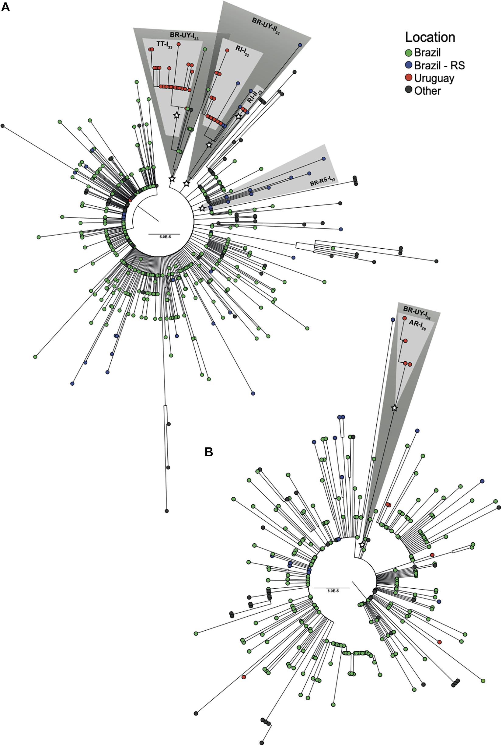
Figure 2. Identification of major SARS-CoV-2 Uruguayan–Brazilian clades. ML phylogenetic trees of (A) 86 B.1.1.33 and (B) 23 B.1.1.28 genomes obtained in this study along with 492 and 275 worldwide reference sequences of the respective genotypes available in GISAID database. Tip circles are colored according to the sampling location. Node support (aLRT) values at key nodes are represented by asterisks (*). Shaded boxes highlight the position of clusters BR-UY-I33, BR-UY-II33, BR-RS-I33, BR-UY-I28, TT-I33, RI-I33, RI-II33, and AR-I28. The tree was rooted on midpoint and branch lengths are drawn to scale with the bars at the center indicating nucleotide substitutions per site. UY-AR, Artigas-Uruguay; UY-RI, Rivera-Uruguay; UY-TT, Treinta y Tres-Uruguay; BR-SE, Southeast Brazilian region; BR-RS, Rio Grande do Sul- Brazil.
The four major Uruguayan clades were nested among basal sequences from Brazil, forming three highly supported (aLRT = 1) Brazilian–Uruguayan clades (Figure 2). The largest clade (n = 39) designated as BR-UY-I33 comprises the Uruguayan clade TT-I33, a group of basal Brazilian sequences (n = 10) isolated in São Paulo and Rio de Janeiro states between March 24 and June 6 and one sequence from Ireland. The second Brazilian–Uruguayan clade designated as BR-UY-II33 comprises the Uruguayan clades RI-I33 (n = 19) and RI-II33 (n = 3) and a group of Brazilian sequences (n = 8) isolated in different municipalities from RS between May 6 and July 28 (Figure 2A). The third Brazilian–Uruguayan clade designated as BR-UY-I28 comprises the Uruguayan clade AR-I28 and one Brazilian sequence isolated in the São Paulo state on March 20 (Figure 2B). Our analysis also identified one highly supported (aLRT = 1) monophyletic group designated as BR-RS-I33 (n = 12) that only comprises sequences from RS (Figure 2A).
Spatiotemporal Dissemination of SARS-CoV-2 Uruguayan–Brazilian Clades
To identify the number of independent introduction events of SARS-CoV-2 into Uruguay and their most probable source location, we used a ML-based probabilistic method of ancestral character state reconstruction implemented in the PastML program. Sequences were grouped according to country (Argentina, Brazil, Chile, and Uruguay) or region (North America, Europe, and Oceania) of origin. The ML phylogeographic analyses estimate nine separate viral introductions into Uruguay from Brazil: five of the lineage B.1.1.28 and four of the lineage B.1.1.33 (Figure 3). Three introductions of lineage B.1.1.33 and one introduction of lineage B.1.1.28 led to onward transmission to more than one individual and gave origin to the Uruguayan clades RI-I33, RI-II33, TT-I33, and AR-I28.
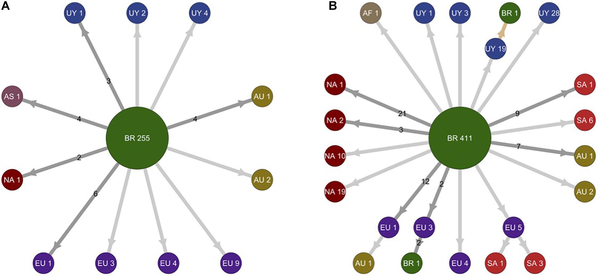
Figure 3. Schematic representation of migration events during worldwide dissemination of SARS-CoV-2 lineages B.1.1.28 and B.1.1.33. The picture depicts the migration events in SARS-CoV-2 lineages B.1.1.28 (A) (n = 298 sequences) and B.1.1.33 (B) (n = 578 sequences) inferred by ancestral character reconstruction obtained through a maximum likelihood (ML) method implemented in PastML. Each node in the network is identified by location and number of sequences within different phylogenetic subclusters. Arrows indicate migration events deduced from location state changes across both trees (B.1.1.28 and B.1.1.33). Dark gray arrows identify multiple migration events and numbers in the arrows quantify the number of migration events connecting respective locations. Light gray arrows identify unique migration events. The light brown arrow points out to a single inferred migration event from Uruguay to Brazil. Nodes are colored according to their location. AF, Africa; AS, Asia; AU, Australia; BR, Brazil; EU, Europe; NA, North America; SA, South America; UY, Uruguay.
To more accurately infer the geographic source and timing of virus introductions in Uruguay, we conducted Bayesian-based phylogeographic and molecular clock analyses of Brazilian and Uruguayan sequences. Viral migrations were inferred using a discrete diffusion model between 13 locations: four Brazilian regions (Southeast, Northeast, North, and Central-West), three southern Brazilian states (RS, Paraná, and Santa Catarina), and six Uruguayan locations (AR, CL, MO, TT, RI, and RO). Bayesian analyses recovered the same Uruguayan and Brazilian clades previously detected by the ML analyses, with two exceptions: the clade RI-II33 branched outside the clade BR-UY-II33 and no Brazilian sequences branched with high support with clade AR-II28. Bayesian analyses pointed that the Brazilian southeast region was the most probable source [posterior state probability (PSP) ≥ 0.96] of all B.1.1.28 and B.1.1.33 viruses introduced in Uruguay, with the exception of clade RI-I33, a B.1.1.28 sequence from RI that most probably originated in Brazil-RS (PSP ≥ 0.85), and the B.1.1.33 strain from Montevideo that probably originated in the North Brazilian region (PSP = 1) (Figure 4 and Supplementary Figures 3, 4).
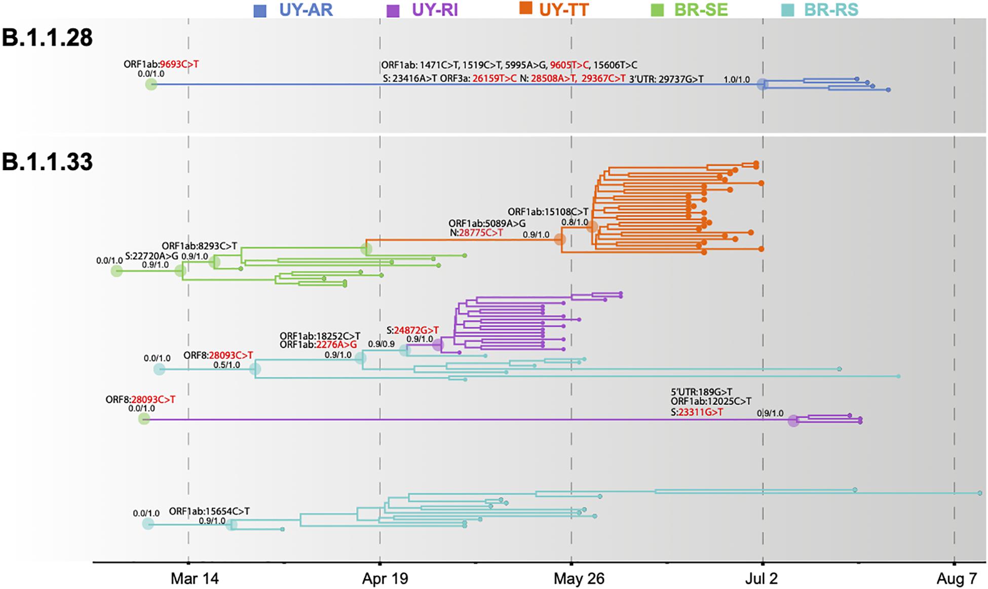
Figure 4. Spatiotemporal dissemination of SARS-CoV-2 Uruguayan–Brazilian clades. Uruguayan–Brazilian clades inferred on the time-scaled Bayesian phylogeographic MCC tree (Supplementary Figures 3, 4) plotted independently. Branches are colored according to the most probable location state of their descendant nodes as indicated at the legend. Posterior probability/posterior state probability support values are indicated at key nodes. Synonymous (black) and non-synonymous (red) substitutions fixed at ancestral nodes are shown.
The temporal origin of the lineages B.1.1.28 and B.1.1.33 was traced back to February 2020 and the TMRCA of major Brazilian–Uruguayan clades were inferred at the following: March 12 (95% HPD: March 09–21) for clade BR-UY-I33, March 23 (95% HPD: March 14–31) for clade BR-RS-I33, March 29 (95% HPD: March 12–April 13) for clade BR-UY-II33, May 2 (95% HPD: April 27–May 04) for clade RI-I33, May 26 (95% HPD: May 12–June 07) for clade TT-I33, July 10 (95% HPD: June 27–July 18) for clade RI-II33, and July 2 (95% HPD: June 17–July 16) for clade AR-I28. The detection lag (time interval between cluster TMRCA and the first detected case) of major B.1.1.33 Uruguayan outbreaks was estimated at 3 days (95% HPD: 1–8 days) for clade RI-I33 and 23 days (95% HPD: 11–37 days) for clade TT-I33. The control lag (time interval between the first detected case and the last transmission event) was estimated at 25 days (95% HPD: 20–28 days) for clade RI-I33 and 10 days (95% HPD: 6–12 days) for clade TT-I33. Though we obtained large confidence intervals, it is worth emphasizing that we were able to assess a remarkable 30% of each outbreak. A larger number of samples might not solve the issue (see discussion on cryptic circulation).
SARS-CoV-2 Lineage-Defining SNPs and Intra-Patient Viral Diversity
Severe acute respiratory syndrome coronavirus 2 Brazilian–Uruguayan clades here identified displayed several lineage-defining SNPs (single nucleotide polymorphisms/mutations) that were sequentially fixed during evolution and dissemination of each lineage (Figure 4 and Supplementary Table 3 in Supplementary Material Section 2). The Uruguayan/Brazilian lineages (BR-UY-I33, BR-UY-II33, and BR-UY-I28) and the Brazilian lineage BR-RS-I33 were all defined by one SNP. One SNP also characterized all sequences from clade TT-I33 plus some basal Brazilian strains from São Paulo, two SNPs defined clade TT-I33, and one additional SNP characterized all except one basal sequence of clade TT-I33. Two SNPs characterized all sequences from clade RI-I33 plus some basal Brazilian strains from RS, one SNP defined clade RI-I33, and three SNPs defined clade RI-II33. The clade AR-I28 was defined by 10 SNPs (Supplementary Table 3 in Supplementary Material Section 2).
To understand the dynamics of lineage-defining SNPs, we examined within-host diversity patterns by analyzing the mapped reads at every SNP genomic position in samples from the two major B.1.1.33 Brazilian–Uruguayan clades (Supplementary Tables 4, 5 in Supplementary Material Section 2). Our analysis of SNP positions reveals a clear-cut allocation of mutant alleles, which displayed either very low (<10%) or high (>80%) frequency within each clade (Figures 5A,B). Importantly, no sequences appeared in the 10–65% frequency range (Figures 5A,B). The most notable example was the mutation ORF1ab:15108C > T fixed during the dissemination of clade TT-I33 that appeared at a very low frequency (<5%) in the basal Uruguayan sample M70 (Figure 5A). Indeed, the estimated frequency of the mutant allele in basal sequences from a given clade was not higher than the corresponding frequency detected in sequences from a different clade (Figure 5C). While allele frequencies were calculated from three different technologies (Illumina, Ion Torrent, and ONT) and this could introduce some bias (Supplementary Figures 1, 2 in Supplementary Material Section 3), for most samples only ONT data were available. It has been shown that in our conditions (we only kept for analyses those synapomorphic sites with at least 100× read coverage), ONT sequencing enables detection of within-specimen single-nucleotide variants at frequencies equal to and higher than ∼40% with adequate accuracy (Bull et al., 2020). Besides, considering intermediate allele frequencies as those in 40–60% range (Bull et al., 2020), small differences in accuracy and precision would not change our main finding regarding the absence of sequence with intermediate frequencies in this dataset.
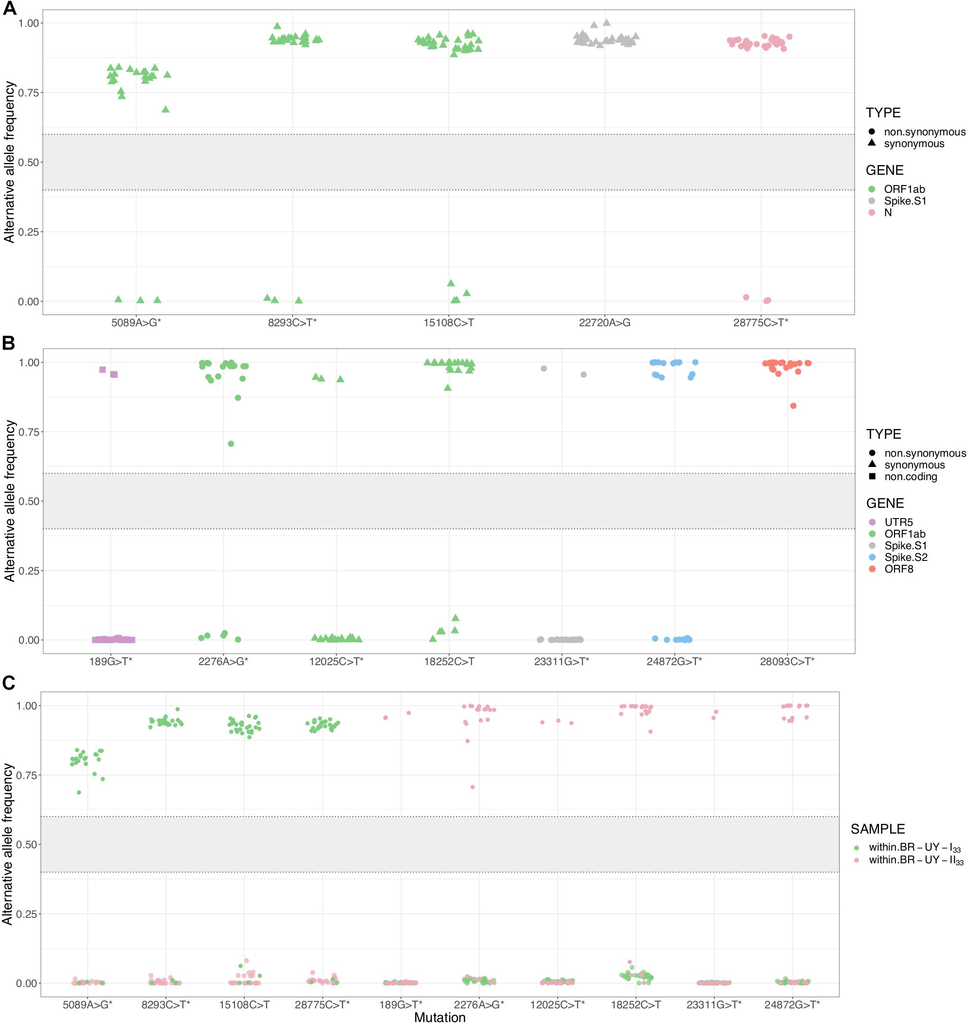
Figure 5. Within-host diversity in synapomorphic sites for the SARS-CoV-2 clades BR-UY-I33 and BR-UY-II33. (A) Alternative allele frequencies for the clade BR-UY-I33, as observed from trimmed bam files. Positions and annotations follow Wuhan’s reference sequence MN908947.3; corresponding gene annotation and mutation types are indicated by color and shape, respectively. The asterisks indicate the synapomorphic sites where the MUTATION explanatory variable shows a significant coefficient in the implemented linear model (p-value ≤ 0.05). In other words, the allele frequencies for these sites are robust to sequencing technology. (B) Same as (A), but for the clade BR-UY-II33. For both clades, the absence of alleles with intermediate frequencies (0.4 < f < 0.6) is notorious. (C) Alternative allele frequencies for all synapomorphies within both BR-UY-I33 and BR-UY-II33 clades. For each mutation, there is an overlap in the alternative allele frequencies of those samples that do not show the synapomorphy, regardless of the clade they belong to. The gray zone in the plot shows the intermediate allele frequency range (40–60%) as defined by Bull et al. (2020).
Discussion
Public health measures implemented by authorities were able to contain the early local expansion of SARS-CoV-2 in Uruguay, but the intensive human mobility across the contiguous dry borderland with Brazil can compromise long-term control of the epidemic. Indeed, our analyses showed that the COVID-19 epidemic in the Uruguayan–Brazilian border is mostly dominated by the Brazilian lineages B.1.1.33 and B.1.1.28 and that independent introductions of these lineages seeded the early outbreaks of SARS-CoV-2 detected in Uruguayan departments at the bordering region.
Our phylogeographic analyses support that both SARS-CoV-2 lineages most probably arose in the Southeast Brazilian region (PSP = 1) during February 2020, consistent with previous findings (Candido et al., 2020; Resende et al., 2020c), and were later disseminated at multiple times into the Southern region, making up 79% of all viral strains from the RS state. We distinguished two B.1.1.33 local sub-clades that together comprise 45% of all B.1.1.33 strains from RS: the clade BR-RS-I33 that arose by the end of March seems to have remained restricted to this Brazilian state, and the clade BR-UY-II33 that arose between late March and early April was later disseminated to Uruguay. These findings indicate that the COVID-19 epidemic in the Southern Brazilian state of RS was mostly seeded by epidemics from the Southeastern region.
The molecular epidemiologic profile of the COVID-19 epidemic at Uruguayan border localities (dominated by lineages B.1.1.33 and B.1.1.28) coincides with the observed pattern in RS state and differs from the profile of early outbreaks in the capital city of Montevideo (dominated by lineages A.5 and B.1) (Elizondo et al., 2020; Salazar et al., 2020). Our phylogeographic analyses support that the Southeast Brazilian region and RS were the most probable sources of six and two viral introductions, respectively, into Uruguay. This finding, however, should be interpreted with caution because a potential sampling bias across Brazilian regions might influence phylogeographic reconstructions and the actual number of viral introductions from RS into Uruguay might be higher than here estimated (Lemey et al., 2020).
Our study supports that some of the viral strains introduced in the Uruguayan cities of Rivera, Treinta y Tres, and Bella Unión between early May and mid-July were able to spread locally. The most successfully disseminated local lineages were RI-I33 and TT-I33 that drove the major early outbreaks detected in Rivera and Treinta y Tres in May and June, respectively. Our analysis supports that SARS-CoV-2 Uruguayan clades displayed a variable period of local cryptic transmission, lasting between 1 and 3 weeks. Furthermore, the major Uruguayan clades took between 10 days and nearly a month to be controlled. Long periods of cryptic circulation and/or long times to control represent a potential risk of viral dissemination from Uruguayan border localities into the metropolitan region.
Our phylogenetic analyses identified several sub-clusters inside the B.1.1.28 and B.1.1.33 lineages defined by a set of synonymous and non-synonymous mutations with respect to the ancestral viruses that arose in Brazil in February 2020. The number of defining mutations ranges from one to 11 SNPs and correlates with the lineage TMRCA, as lineages with the youngest TMRCA displayed more SNPs than those with older ones. This is consistent with a molecular clock model (Duchene et al., 2020) and with the notion that most SNPs are probably evolutionarily neutral (van Dorp et al., 2020). Thus, while our study emphasizes the rapid genetic differentiation of regional SARS-CoV-2 lineages in South America and the relevance of molecular epidemiologic studies to track their emergence and spread, the observed SNPs should not be interpreted as an adaptive signature of higher viral transmissibility (MacLean et al., 2020).
Previous studies support that intra-host SARS-CoV-2 genetic diversity is transmissible, and information about within-host diversity could provide further resolution for identification of geographic transmission clusters (Lythgoe et al., 2020) and to trace how low-frequency mutations become fixed in local clusters (Popa et al., 2020). Our analyses of intra-host diversity, however, revealed that mutations present at low frequency (<10%) in basal strains of major Uruguayan–Brazilian clusters become highly prevalent (>80%) during subsequent transmissions, without evidence of basal samples with intermediate-frequency alternative alleles. These findings support that minor intra-host SARS-CoV-2 variants could become rapidly fixed due to stochastic events during inter-host transmissions and later perpetuated along transmission chains (Böhmer et al., 2020).
In summary, our study demonstrates for the first time a recurrent viral flux across the Uruguayan–Brazilian border, with multiple introductions of the SARS-CoV-2 Brazilian lineages B.1.1.28 and B.1.1.33 into Uruguayan localities at the border region and the potential of those viral introductions to ignite local outbreaks in Uruguay. These findings provide clear evidence that public health measures for viral control were not able to fully suppress the dissemination of SARS-CoV-2 along the Uruguayan-Brazilian border, and this possesses a major challenge for long-term epidemic control in Uruguay. The present work also highlights the relevance of bi-national consortiums for genomic surveillance of SARS-CoV-2 to obtain in-depth insights and better control of viral spread across highly permissive international borders.
Data Availability Statement
Genome sequences were submitted to the EpiCoV/GISAID database, and accession numbers are specified in Supplementary Tables 1, 2 and in Supplementary Section 2.
Ethics Statement
The studies involving human participants were reviewed and approved by the SASA Ethics Committee: CEI-SASA FIOCRUZ-IOC Ethics Committee: 68118417.6.0000.5248, and Brazilian Ministry of Health SISGEN: A1767C3. The patients/participants provided their written informed consent to participate in this study.
Author Contributions
LS made significant contributions in the post-processing of raw sequencing data, study design, coordination of Uruguayan group, funding acquisition, verifying underlying data, and writing the original draft. DM was in charge of phylogenetic and phylogeographic analyses, verified underlying data, and wrote original draft. NRg made significant contribution in the post-processing of raw sequencing data, verifying underlying data, variant analysis, and writing the original draft. TF-C was in charge of sequencing in Uruguay and read and corrected the manuscript. VN is the head of a diagnostic center in the Sanatorio Americano, obtained funding, and reviewed and edited the manuscript. MA, TP, and NRy worked on the diagnosis in Sanatorio Americano and read the manuscript. GB made significant contributions in the study design, coordination between Uruguayan and Brazilian institutions, coordination of phylogeographic and phylogenetic analyses, verification of underlying data, and writing the original draft. RC made significant contributions in the coordination of diagnostic centers in the Uruguayan non-metropolitan area, study design, and editing and review of the manuscript. FT worked on the diagnosis in CENUR-Norte, Uruguay, worked on nanopore sequencing in Uruguay centers, and edited and reviewed the manuscript. MB worked on nanopore sequencing in Uruguay centers and read the manuscript. MG made significant contributions in the technical support at the Institut Pasteur de Montevideo, helped in writing the grant for funding, and carefully edited and reviewed the manuscript. CSa made significant contributions in the bioinformatic analysis of sequencing data from Uruguayan centers and reviewed the manuscript. MV, AL, MC, LMay, and MSa worked on diagnosis in CENUR-Norte, Uruguay, and reviewed the manuscript. YV worked on diagnosis in Laboratorio DILAVE/MGAP-INIA-UdelaR-Tacuarembó, Uruguay, and reviewed the manuscript. CA worked on diagnosis in CURE-Rocha, Uruguay, and reviewed the manuscript. CSe worked on nanopore sequencing at Institut Pasteur de Montevideo, Uruguay, and reviewed the manuscript. IF made significant contributions in the software and reviewed the manuscript. PS and JS worked on Ion Torrent sequencing at Instituto de Investigaciones Biológicas Clemente Estable, Uruguay, and reviewed the manuscript. RF and CM made significant contributions in sequencing design, library preparation, and sequencing (Ion Torrent). MB-G worked on logistic support and reviewed the manuscript. IA worked on phylogenetic and phylogeographic analyses and reviewed the manuscript. PR coordinated the Brazilian SARS-CoV-2 sequencing team using Nanopore, Illumina, and Ion Torrent platforms at the National Reference Laboratory Fiocruz; analyzed the raw sequencing data; and revised and edited the manuscript. TG and MM performed the extraction and the SARS-CoV-2 detection by real-time RT-PCR locally in the Rio Grande do Sul state and revised the manuscript. LMar is responsible for the epidemiological team in the Rio Grande do Sul state, collected all the epidemiological data from Brazilian samples included in this study, and revised the manuscript. FM is responsible for extraction and the SARS-CoV-2 confirmation by real-time RT-PCR at the National Reference Laboratory Fiocruz and revised the manuscript. LA and AM are responsible for whole-genome SARS-CoV-2 amplification, library construction, sequencing, and revision of the manuscript. MMS made significant contribution in financial support acquisition and revision and editing of the manuscript. All authors contributed to the article and approved the submitted version.
Funding
Funding support in Brazil was from CGLab/MoH (General Laboratories Coordination of Brazilian Ministry of Health), CVSLR/FIOCRUZ (Coordination of Health Surveillance and Reference Laboratories of Oswaldo Cruz Foundation), CNPq COVID-19 MCTI 402457/2020-0, and INOVA VPPCB-005-FIO437 20-2. In Uruguay, this work was funded by the Manuel Perez Foundation that nucleated COVID-19 donation funds in the context of the project “Vigilancia epidemiológica del COVID-19 en las fronteras uruguayas y ana lisis de su transmisi n en el interior del pai s.” This work was also supported by FOCEM – Fondo para la Convergencia Estructural del Mercosur (COF 03/11). GB was funded by productivity research fellowships from the Conselho Nacional de Desenvolvimento Cient fico e Tecnol gico – CNPq (Grant No. 302317/2017-1) and the Funda o Carlos Chagas Filho de Amparo Pesquisa do Estado do Rio de Janeiro – FAPERJ (Grant No. E-26/202.896/2018). Members of the Institut Pasteur de Montevideo were supported by FOCEM-Fondo para la Convergencia Estructural del Mercosur (COF03/11).
Conflict of Interest
The authors declare that the research was conducted in the absence of any commercial or financial relationships that could be construed as a potential conflict of interest.
Acknowledgments
We thank to the kind donors for their contribution to this project. We thank to Carolina Banchero and Rosina Segui for beautiful figure design. We thank to Hugo Naya for helping with statistical models and Luisa Berna for fruitful discussions. We are grateful to the Laboratory of Experimental Evolution of Viruses in charge Gonzalo Moratorio, the Laboratory of Microbial Genomics in charge Gregorio Iraola, the Unit of Molecular Biology in charge Carlos Robello, and all of the people from the Institut Pasteur de Montevideo, for making their facilities available at any time for our experiments and sharing their experience, as well as equipment, plastic supplies, and reagents. We thank Gabriela Ortiz and Paula Aguerrebere of the IAC (Instituto Asistencial Colectivo, Treinta y Tres) for their willingness and hard work in contacting patients. We also thank the workforce of the technical group of the Laboratory for Respiratory Viruses at LACEN/CEVS/SES-RS and the support of the Center for Scientific and Technological Development (CDCT/CEVS/SES-RS). Finally, we thank for efforts from different groups that contribute with SARS-CoV-2 genomes to the EpiCoV GISAID initiative (Supplementary Tables 9, 10 in Supplementary Material Section 2).
Supplementary Material
The Supplementary Material for this article can be found online at: https://www.frontiersin.org/articles/10.3389/fmicb.2021.653986/full#supplementary-material
Footnotes
References
Anisimova, M., and Gascuel, O. (2006). Approximate likelihood-ratio test for branches: A fast, accurate, and powerful alternative. Syst. Biol. 55, 539–552. doi: 10.1080/10635150600755453
Arnson, C. J., Gedan, B., and Prusa, A. (2020). Contagion: Brazil’s COVID-19 Catastrophe Spills Over Borders. Washington, DC: Wilson Center. Available online at: https://www.wilsoncenter.org/blog-post/contagion-brazils-covid-19-catastrophe-spills-over-borders (accessed december 2, 2020)
Böhmer, M. M., Buchholz, U., Corman, V. M., Hoch, M., Katz, K., Marosevic, D., et al. (2020). Articles Investigation of a COVID-19 outbreak in Germany resulting from a single travel-associated primary case: a case series. Lancet Infect. Dis. 20, 920–928. doi: 10.1016/S1473-3099(20)30314-5
Bull, R. A., Adikari, T. N., Ferguson, J. M., Hammond, J. M., Stevanovski, I., Beukers, A. G., et al. (2020). Analytical validity of nanopore sequencing for rapid SARS-CoV-2 genome analysis. Nat. Commun. 11:6272. doi: 10.1038/s41467-020-20075-6
Candido, D. S., Claro, I. M., de Jesus, J. G., Souza, W. M., Moreira, F. R. R., Dellicour, S., et al. (2020). Evolution and epidemic spread of SARS-CoV-2 in Brazil. Science 369, 1255–1260. doi: 10.1126/science.abd2161
Duchene, S., Featherstone, L., Haritopoulou-Sinanidou, M., Rambaut, A., Lemey, P., and Baele, G. (2020). Temporal signal and the phylodynamic threshold of SARS-CoV-2. bioRxiv 077735. [pre-print]. doi: 10.1101/2020.05.04.077735
Elizondo, V., Harkins, G. W., Mabvakure, B., Smidt, S., Zappile, P., Marier, C., et al. (2020). SARS-CoV-2 genomic characterization and clinical manifestation of the COVID-19 outbreak in Uruguay. Emerg. Microb. Infect. 2020:1863747. doi: 10.1080/22221751.2020.1863747
Ferreira, M., and Suchard, M. A. (2008). Bayesian analysis of elapsed times in continuous-time Markov chains. Canad. J. Statist. 36:5550360302. doi: 10.1002/cjs.5550360302
github (2020). Estadísticas epidemiológicas de Uruguay. San Francisco: github. Available online at: https://guiad-covid.github.io/data/estadisticasuy/. (accessed december 2, 2020)
Guindon, S., Dufayard, J. F., Lefort, V., Anisimova, M., Hordijk, W., and Gascuel, O. (2010). New algorithms and methods to estimate maximum-likelihood phylogenies: assessing the performance of PhyML 3.0. Syst. Biol. 59, 307–321. doi: 10.1093/sysbio/syq010
Guindon, S., Lethiec, F., Duroux, P., and Gascuel, O. (2005). PHYML Online–a web server for fast maximum likelihood-based phylogenetic inference. Nucleic Acids Res. 33, W557–W559. doi: 10.1093/nar/gki352
Ishikawa, S., Zhukova, A., Iwasaki, W., and Gascuel, O. (2019). A Fast Likelihood Method to Reconstruct and Visualize Ancestral Scenarios. Mol. Biol. Evolut. 36, 2069–2085. doi: 10.1093/molbev/msz131
Lemey, P., Hong, S. L., Hill, V., Baele, G., Poletto, C., Colizza, V., et al. (2020). Accommodating individual travel history and unsampled diversity in Bayesian phylogeographic inference of SARS-CoV-2. Nat. Commun. 11:5110. doi: 10.1038/s41467-020-18877-9
Lythgoe, K. A., Hall, M., Ferretti, L., de Cesare, M., MacIntyre-Cockett, G., Trebes, A., et al. (2020). Within-host genomics of SARS-CoV-2. bioRxiv 118992. [preprint]. doi: 10.1101/2020.05.28.118992
MacLean, O. A., Orton, R. J., Singer, J. B., and Robertson, D. L. (2020). No evidence for distinct types in the evolution of SARS-CoV-2. Virus Evolut. 6:veaa034. doi: 10.1093/ve/veaa034
Moreno, P., Moratorio, G., Iraola, G., Fajardo, A., Aldunate, F., Pereira, M., et al. (2020). An effective COVID-19 response in South America: the Uruguayan Conundrum. medRxiv [preprint]. doi: 10.1101/2020.07.24.20161802
Popa, A., Genger, J. W., Nicholson, M., Penz, T., Schmid, D., Aberle, S. W., et al. (2020). Mutational dynamics and transmission properties of SARS-CoV-2 superspreading events in Austria. bioRxiv 204339 [preprint]. doi: 10.1101/2020.07.15.204339
Quick, J. (2020). nCoV-2019 sequencing protocol V.1. Protocols 2020:bbmuik6w. doi: 10.17504/protocols.io.bbmuik6w
Rambaut, A., Holmes, E. C., O’Toole, Á, Hill, V., McCrone, J. T., Ruis, C., et al. (2020). A dynamic nomenclature proposal for SARS-CoV-2 lineages to assist genomic epidemiology. Nat. Microbiol. 5, 1403–1407. doi: 10.1038/s41564-020-0770-5
Resende, P. (2020). Long reads nanopore sequencing to recover SARS-CoV-2 whole genome V.3. Protocols 2020:bfy7jzn. doi: 10.17504/protocols.io.bfy7jpzn
Resende, P. C., Couto Motta, F., Roy, S., Appolinario, L., Fabri, A., Xavier, J., et al. (2020a). SARS-CoV-2 genomes recovered by long amplicon tiling multiplex approach using nanopore sequencing and applicable to other sequencing platforms. bioRxiv 069039. [preprint]. doi: 10.1101/2020.04.30.069039
Resende, P. C., Delatorre, E., Gräf, T., Mir, D., Couto Motta, F., Appolinario, L., et al. (2020c). Genomic surveillance of SARS-CoV-2 reveals community transmission of a major lineage during the early pandemic phase in Brazil. bioRxiv 2020:158006. doi: 10.1101/2020.06.17.158006
Resende, P. C., Delatorre, E., Gräf, T., Mir, D., Motta, F., and Appolinario, L. (2020b). Evolutionary dynamics and dissemination pattern of the SARS-CoV-2 lineage B.1.1.33 during the early pandemic phase in Brazil. Front. Microbiol. 11:615280. doi: 10.3389/fmicb.2020.615280
Salazar, C., Díaz-Viraqué, F., Pereira-Gómez, M., Ferrés, I., Moreno, P., Moratorio, G., et al. (2020). Multiple introductions, regional spread and local differentiation during the first week of COVID-19 epidemic in Montevideo, Uruguay. bioRxiv 086223 [preprint]. doi: 10.1101/2020.05.09.086223
saude (2021). Painel Coronavírus RS. Available online at: https://ti.saude.rs.gov.br/covid19/ (accessed April 30, 2021).
Suchard, M. A., Lemey, P., Baele, G., Ayres, D. L., Drummond, A. J., and Rambaut, A. (2018). Bayesian phylogenetic and phylodynamic data integration using BEAST 1.10. Virus Evolut. 4:1000520. doi: 10.1371/journal.pcbi.1000520
Valcarcel, B., Avilez, J. L., Torres-Roman, J. S., Poterico, J. A., Bazalar-Palacios, J., La Vecchia, C., et al. (2020). The effect of early-stage public health policies in the transmission of COVID-19 for South American countries. Rev. Panam Salud Publica. 44:e148. doi: 10.26633/RPSP.2020.148
van Dorp, L., Richard, D., Tan, C. C. S., Shaw, L. P., Acman, M., and Balloux, F. (2020). No evidence for increased transmissibility from recurrent mutations in SARS-CoV-2. Nat. Commun. 11:5986. doi: 10.1038/s41467-020-19818-2
Worldometers (2020). COVID-19 CORONAVIRUS PANDEMIC. Wuhan: Worldometers. Available online at: https://www.worldometers.info/coronavirus/ (accessed december 15, 2020)
Keywords: genomics, epidemiology, phylogeography, phylogenetics, SARS-CoV-2, Uruguay, Brazil
Citation: Mir D, Rego N, Resende PC, Tort F, Fernández-Calero T, Noya V, Brandes M, Possi T, Arleo M, Reyes N, Victoria M, Lizasoain A, Castells M, Maya L, Salvo M, Schäffer Gregianini T, Mar da Rosa MT, Garay Martins L, Alonso C, Vega Y, Salazar C, Ferrés I, Smircich P, Sotelo Silveira J, Fort RS, Mathó C, Arantes I, Appolinario L, Mendonça AC, Benítez-Galeano MJ, Simoes C, Graña M, Motta F, Siqueira MM, Bello G, Colina R and Spangenberg L (2021) Recurrent Dissemination of SARS-CoV-2 Through the Uruguayan–Brazilian Border. Front. Microbiol. 12:653986. doi: 10.3389/fmicb.2021.653986
Received: 15 January 2021; Accepted: 08 April 2021;
Published: 28 May 2021.
Edited by:
Kai Huang, University of Texas Medical Branch, United StatesReviewed by:
Matthew M. Hernandez, Icahn School of Medicine at Mount Sinai, United StatesPiyush Baindara, University of Missouri, United States
Xianding Deng, University of California, San Francisco, United States
Copyright © 2021 Mir, Rego, Resende, Tort, Fernández-Calero, Noya, Brandes, Possi, Arleo, Reyes, Victoria, Lizasoain, Castells, Maya, Salvo, Schäffer Gregianini, Mar da Rosa, Garay Martins, Alonso, Vega, Salazar, Ferrés, Smircich, Sotelo Silveira, Fort, Mathó, Arantes, Appolinario, Mendonça, Benítez-Galeano, Simoes, Graña, Motta, Siqueira, Bello, Colina and Spangenberg. This is an open-access article distributed under the terms of the Creative Commons Attribution License (CC BY). The use, distribution or reproduction in other forums is permitted, provided the original author(s) and the copyright owner(s) are credited and that the original publication in this journal is cited, in accordance with accepted academic practice. No use, distribution or reproduction is permitted which does not comply with these terms.
*Correspondence: Luciía Spangenberg, lucia@pasteur.edu.uy; Rodney Colina, rodneycolina1@gmail.com; Gonzalo Bello, gbellobr@gmail.com
†These authors have contributed equally to this work