
Effects of COVID-19 on Synaptic and Neuronal Degeneration
 , , , , , and
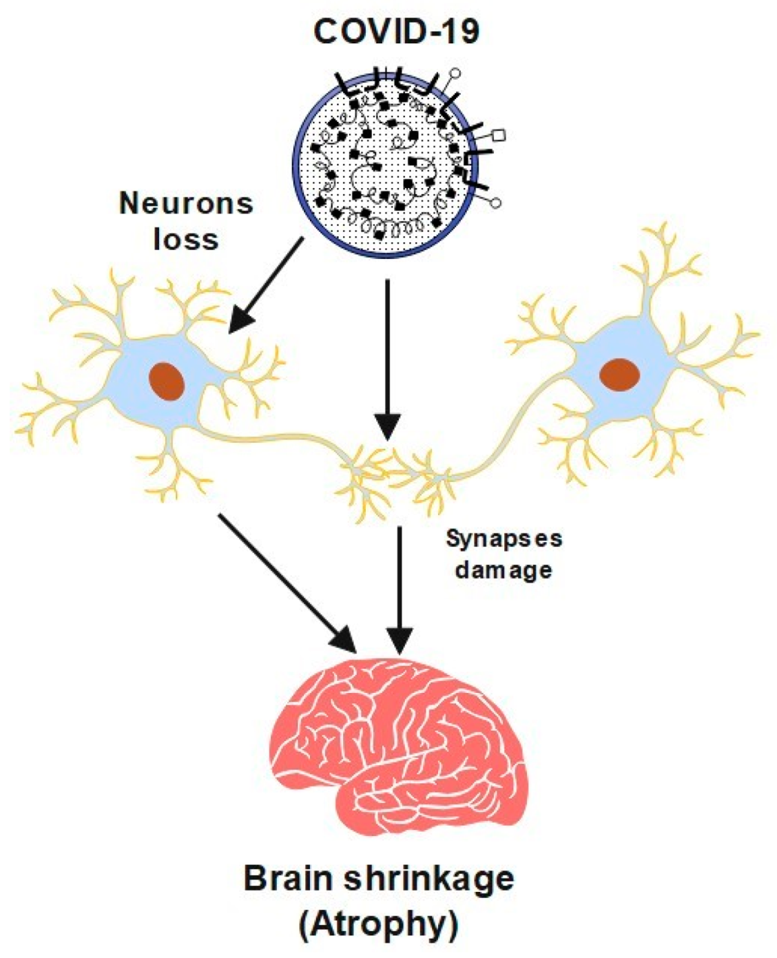
Abstract
:1. Introduction
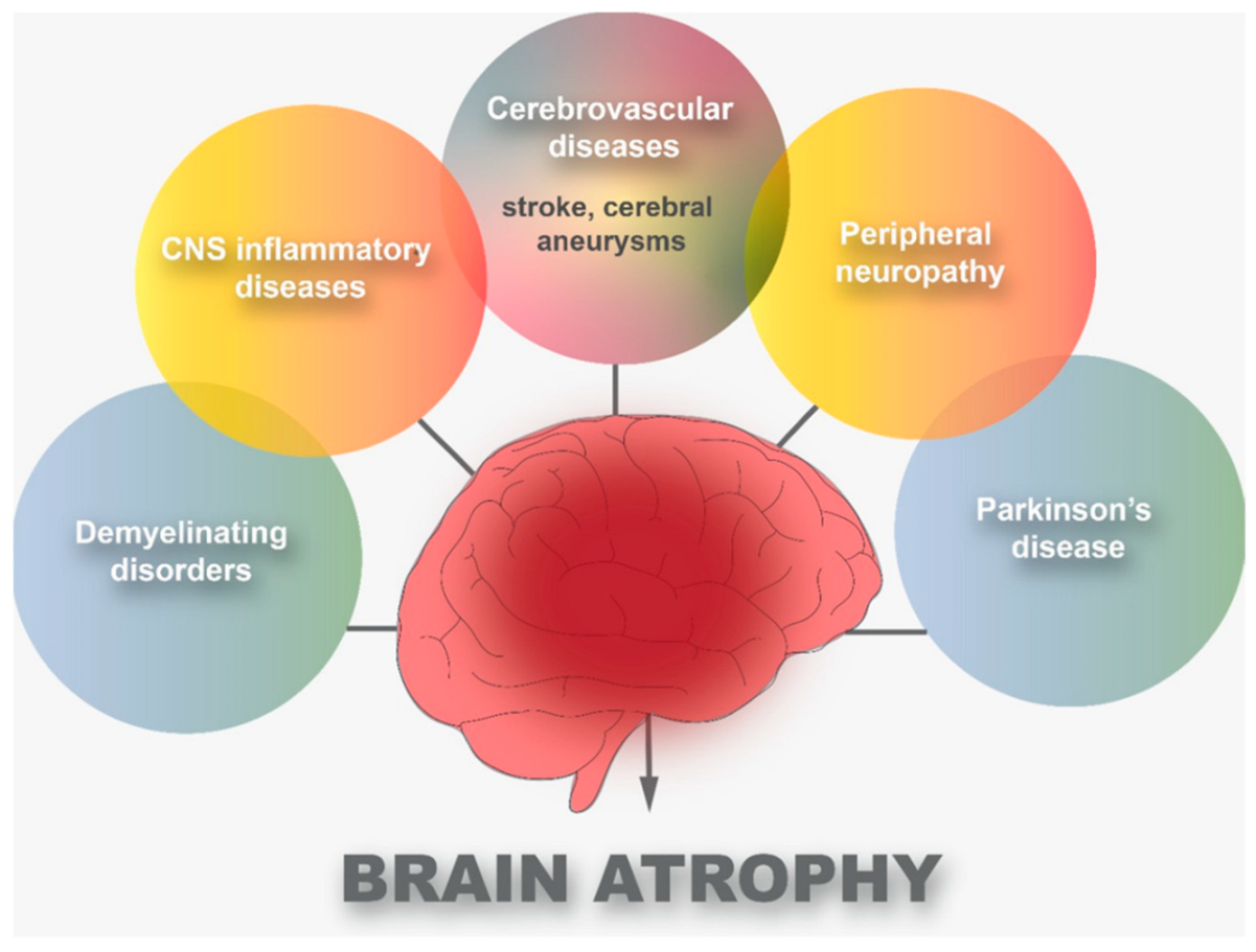
2. Cerebrovascular Diseases and COVID-19
3. Peripheral Neuropathy and CNS Inflammatory Diseases
Peripheral Neuropathy and CNS Inflammatory Diseases
4. COVID-19 and Parkinson’s Disease
COVID-19 and Parkinson’s Disease
5. Fetal and Children Brain Damage
6. Alzheimer’s Disease (AD) and COVID-19
7. COVID-19 Patients with Anosmia
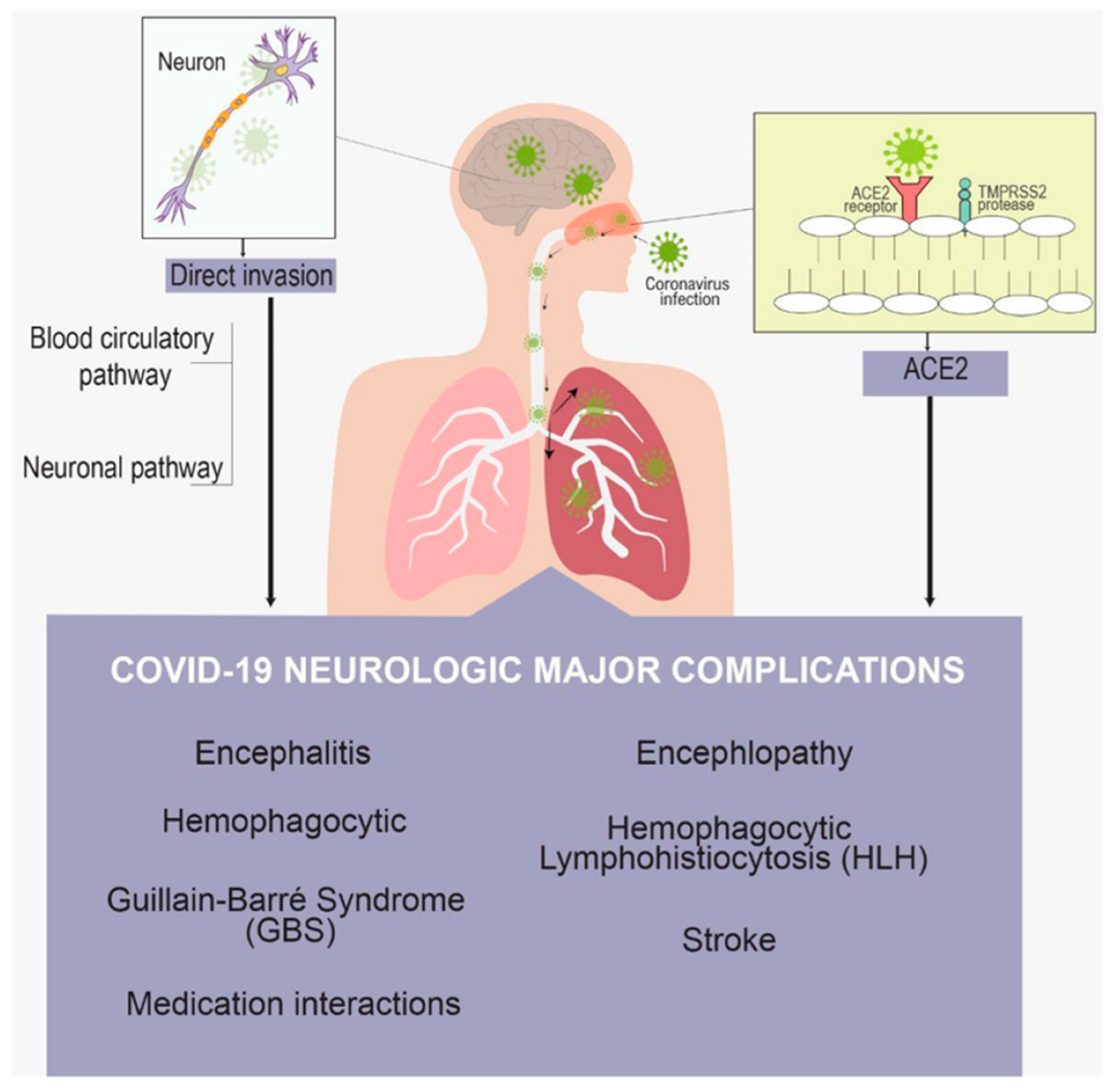
8. Neurological Manifestation
9. Conclusions and Future Perspectives
Author Contributions
Funding
Institutional Review Board Statement
Informed Consent Statement
Data Availability Statement
Acknowledgments
Conflicts of Interest
References
- Verma, K.; Amitabh Prasad, D.N.; Kumar, B.; Kohli, E. Brain and COVID-19 Crosstalk: Pathophysiological and Psychological Manifestations. ACS Chem. Neurosci. 2020, 11, 3194–3203. [Google Scholar] [CrossRef] [PubMed]
- Li, Z.; Liu, T.; Yang, N.; Han, D.; Mi, X.; Li, Y.; Liu, K.; Vuylsteke, A.; Xiang, H.; Guo, X. Neurological manifestations of patients with COVID-19: Potential routes of SARS-CoV-2 neuroinvasion from the periphery to the brain. Front. Med. 2020, 14, 533–541. [Google Scholar] [CrossRef] [PubMed]
- Dixon, L.; Varley, J.A.; Gontsarova, A.; Mallon, D.; Tona, F.; Muir, D.A.; Luqmani, A.; Jenkins, I.H.; Nicholas, R.S.; Jones, B.; et al. COVID-19-related acute necrotizing encephalopathy with brain stem involvement in a patient with aplastic anemia. Neurol. Neuroimmunol. Neuroinflammation 2020, 7, e789. [Google Scholar] [CrossRef] [PubMed]
- Coolen, T.; Lolli, V.; Sadeghi, N.; Rovai, A.; Trotta, N.; Taccone, F.S.; Creteur, J.; Henrard, S.; Goffard, J.; Dewitte, O.; et al. Early postmortem brain MRI findings in COVID-19 non-survivors. Neurology 2020, 95, e2016-27. [Google Scholar] [CrossRef] [PubMed]
- Gandhi, S.; Srivastava, A.K.; Ray, U.; Tripathi, P.P. Is the Collapse of the Respiratory Center in the Brain Responsible for Respiratory Breakdown in COVID-19 Patients? ACS Chem. Neurosci. 2020, 11, 1379–1381. [Google Scholar] [CrossRef]
- Espinosa, P.S.; Rizvi, Z.; Sharma, P.; Hindi, F.; Filatov, A. Neurological Complications of Coronavirus Disease (COVID-19): Encephalopathy, MRI Brain and Cerebrospinal Fluid Findings: Case 2. Cureus 2020, 12, e7930. [Google Scholar] [CrossRef]
- Hagerty, S.L.; Williams, L.M. The impact of COVID-19 on mental health: The interactive roles of brain biotypes and human connection. Brain Behav. Immun. Health 2020, 5, 100078. [Google Scholar] [CrossRef]
- Yin, X.; Zheng, X.; Peng, W.; Wu, M.; Mao, X. Vascular Endothelial Growth Factor (VEGF) as a Vital Target for Brain Inflammation during the COVID-19 Outbreak. ACS Chem. Neurosci. 2020, 11, 1704–1705. [Google Scholar] [CrossRef]
- Lin, E.; Lantos, J.; Strauss, S.; Phillips, C.; Campion, T.R.; Navi, B.B.; Parikh, N.S.; Merkler, A.E.; Mir, S.; Zhang, C.; et al. Brain Imaging of Patients with COVID-19: Findings at an Academic Institution during the Height of the Outbreak in New York City. Am. J. Neuroradiol. 2020, 41, 2001–2008. [Google Scholar] [CrossRef]
- Chigr, F.; Merzouki, M.; Najimi, M. Autonomic Brain Centers and Pathophysiology of COVID-19. ACS Chem. Neurosci. 2020, 11, 1520–1522. [Google Scholar] [CrossRef]
- Saavedra, J.M. COVID-19, Angiotensin Receptor Blockers, and the Brain. Cell. Mol. Neurobiol. 2020, 40, 667–674. [Google Scholar] [CrossRef] [PubMed]
- Fischer, D.; Threlkeld, Z.D.; Bodien, Y.G.; Kirsch, J.E.; Huang, S.; Schaefer, P.W.; Rapalino, O.; Hochberg, L.R.; Rosen, B.R.; Edlow, B.L. Intact Brain Network Function in an Unresponsive Patient with COVID-19. Ann. Neurol. 2020, 88, 851–854. [Google Scholar] [CrossRef] [PubMed]
- Crunfli, F.; Carregari, V.C.; Veras, F.P.; Vendramini, P.H.; Valença, A.G.; Antunes, A.S.; Brandão-Teles, C.; Zuccoli, G.D.; Reis-de-Oliveira, G.; Silva-Costa, L.C.; et al. SARS-CoV-2 infects brain astrocytes of COVID-19 patients and impairs neuronal viability. medRxiv 2021. [Google Scholar] [CrossRef]
- Achar, A.; Ghosh, C. COVID-19-Associated Neurological Disorders: The Potential Route of CNS Invasion and Blood-Brain Barrier Relevance. Cells 2020, 9, 2360. [Google Scholar] [CrossRef] [PubMed]
- Yang, A.C.; Kern, F.; Losada, P.M.; Maat, C.A.; Schmartz, G.P.; Fehlmann, T.; Schaum, N.; Lee, D.P.; Calcuttawala, K.; Vest, R.T.; et al. Broad transcriptional dysregulation of brain and choroid plexus cell types with COVID-19. BioRxiv 2020. [Google Scholar] [CrossRef]
- Fabbri, V.P.; Foschini, M.P.; Lazzarotto, T.; Gabrielli, L.; Cenacchi, G.; Gallo, C.; Aspide, R.; Frascaroli, G.; Cortelli, P.; Riefolo, M.; et al. Brain ischemic injury in COVID-19-infected patients: A series of 10 post-mortem cases. Brain Pathol. 2020, 31, 205–210. [Google Scholar] [CrossRef]
- Pinggera, D.; Klein, B.E.; Thomė, C.; Grassner, L. The influence of the COVID-19 pandemic on traumatic brain injuries in Tyrol: Experiences from a state under lockdown. Eur. J. Trauma Emerg. Surg. 2020, 47, 653–658. [Google Scholar] [CrossRef] [PubMed]
- Malentacchi, M.; Gned, D.; Angelino, V.; Demichelis, S.; Perboni, A.; Veltri, A.; Bertolotto, A.; Capobianco, M. Concomitant brain arterial and venous thrombosis in a COVID-19 patient. Eur. J. Neurol. 2020, 27, e38–e39. [Google Scholar] [CrossRef]
- Haider, A.; Siddiqa, A.; Ali, N.; Dhallu, M. COVID-19 and the Brain: Acute Encephalitis as a Clinical Manifestation. Cureus 2020, 12, e10784. [Google Scholar] [CrossRef]
- Mao, X.; Jin, W. The COVID-19 Pandemic: Consideration for Brain Infection. Neuroscience 2020, 437, 130–131. [Google Scholar] [CrossRef]
- Choi, J.; Lee, H.; Park, J.; Cho, S.; Kwon, M.; Jo, C.; Koh, Y.H. Altered COVID-19 receptor ACE2 expression in a higher risk group for cerebrovascular disease and ischemic stroke. Biochem. Biophys. Res. Commun. 2020, 528, 413–419. [Google Scholar] [CrossRef] [PubMed]
- Altable, M.; De la Serna, J. Cerebrovascular disease in COVID-19: Is there a higher risk of stroke? Brain Behav. Immun. Health 2020, 6, 100092. [Google Scholar] [CrossRef] [PubMed]
- Escalard, S.; Chalumeau, V.; Escalard, C.; Redjem, H.; Delvoye, F.; Hébert, S.; Smajda, S.; Ciccio, G.; Desilles, J.P.; Mazighi, M.; et al. Early Brain Imaging Shows Increased Severity of Acute Ischemic Strokes with Large Vessel Occlusion in COVID-19 Patients. Stroke 2020, 51, 3366–3370. [Google Scholar] [CrossRef] [PubMed]
- Voznyuk, I.A.; Ilyina, O.; Kolomentsev, S.V. Ischemic Stroke as a Clinical Form and Pathogenetic Model in the Structure of Central Nervous System Lesions in COVID-19. Bull. Rehabil. Med. 2020, 4, 90–98. [Google Scholar] [CrossRef]
- Bekelis, K.; Missios, S.; Ahmad, J.; Labropoulos, N.; Schirmer, C.M.; Calnan, D.R.; Skinner, J.; Mackenzie, T. Ischemic Stroke Occurs Less Frequently in Patients with COVID-19. Stroke 2020, 51, 3570–3576. [Google Scholar] [CrossRef]
- Landa, N.A.; Oficialdegui, C.V.; Fernández, K.I.; Larrabe, I.G.; Onaindia, S.R.; Benguria, S.T. Ischemic-hemorrhagic stroke in patients with Covid-19. Rev. Espanñola Anestesiol. Y Reanim. 2020, 67, 516–520. [Google Scholar]
- Zhou, Y.; Lu, J.; Cheng, Y.; Xin, N. Nervous system complications of COVID-19 with a focus on stroke. Eur. Rev. Med. Pharmacol. Sci. 2020, 24, 13044–13048. [Google Scholar]
- Nunes, N.D.; Nascimento, J.F.; Nascimento, J.K.; Castro, R.R.; Azizi, M.A.; Junior, G.C.; Catharino, A.M.; Silveira, V.; Neves, M.A. Brain and Covid-19: An Integrative Review. E-Cronicon Neurol. 2020, 12, 101–107. [Google Scholar]
- Pennisi, M.; Lanza, G.; Falzone, L.; Fisicaro, F.; Ferri, R.; Bella, R. SARS-CoV-2 and the Nervous System: From Clinical Features to Molecular Mechanisms. Int. J. Mol. Sci. 2020, 21, 5475. [Google Scholar] [CrossRef]
- Goldberg, M.F.; Goldberg, M.F.; Cerejo, R.; Tayal, A. Cerebrovascular Disease in COVID-19. Am. J. Neuroradiol. 2020, 41, 1170–1172. [Google Scholar] [CrossRef]
- Hernández-Fernández, F.; Sandoval Valencia, H.; Barbella-Aponte, R.A.; Collado-Jiménez, R.M.; Ayo-Martin, O.; Barrena, C.; Molina-Nuevo, J.D.; García-García, J.; Lozano-Setién, E.; Alcahut-Rodríguez, C.; et al. Cerebrovascular disease in patients with COVID-19: Neuroimaging, histological and clinical description. Brain 2020, 143, 3089–3103. [Google Scholar] [CrossRef] [PubMed]
- Rea, G.; Lassandro, F.; Lieto, R.; Bocchini, G.; Romano, F.Y.; Sica, G.; Valente, T.; Muto, E.; Murino, P.; Pinto, A.; et al. Lesson by SARS-CoV-2 disease (COVID-19): Whole-body CT angiography detection of “relevant” and “other/incidental” systemic vascular findings. Eur. Radiol. 2021, 31, 7363–7370. [Google Scholar] [CrossRef] [PubMed]
- Bittmann, S. COVID-19: Expression of ACE2-receptors in the Brain Suggest Neurotropic Damage. J. Regen. Biol. Med. 2020, 2, 1–3. [Google Scholar] [CrossRef] [PubMed]
- Jaunmuktane, Z.; Mahadeva, U.; Green, A.C.; Sekhawat, V.; Barrett, N.A.; Childs, L.; Shankar-Hari, M.; Thom, M.; Jäger, H.R.; Brandner, S. Microvascular injury and hypoxic damage: Emerging neuropathological signatures in COVID-19. Acta Neuropathol. 2020, 140, 397–400. [Google Scholar] [CrossRef]
- Boyko, A.N.; Sivertseva, S.; Spirin, N.N. Nervous system damage in COVID-19 with an emphasis on the management of patients with multiple sclerosis. Neurol. Neuropsychiatry Psychosom. 2020, 12, 44–47. [Google Scholar] [CrossRef]
- Mehta, S.; Bhandari, S.; Mehta, S. Brain autopsies in fatal COVID-19 and postulated pathophysiology: More puzzling than a Rubik’s cube. J. Clin. Pathol. 2020, 74, 612–613. [Google Scholar] [CrossRef]
- Babkina, A.S.; Golubev, A.; Ostrova, I.V.; Volkov, A.V.; Kuzovlev, A.N. Brain Morphological Changes in COVID-19. Gen. Reanimatol. 2021, 17, 4–15. [Google Scholar] [CrossRef]
- Haque, E.; Akther, M.; Azam, S.; Kim, I.; Choi, D. COVID-19 and The Brain. J. Microb. Biochem. Technol. 2020, 12, 1–9. [Google Scholar]
- Bulfamante, G.; Bocci, T.; Falleni, M.; Campiglio, L.; Coppola, S.; Tosi, D.; Chiumello, D.; Priori, A. Brainstem neuropathology in two cases of COVID-19: SARS-CoV-2 trafficking between brain and lung. J. Neurol. 2021, 268, 4486–4491. [Google Scholar] [CrossRef]
- Jha, N.K.; Ojha, S.K.; Jha, S.K.; Dureja, H.; Singh, S.K.; Shukla, S.D.; Chellappan, D.K.; Gupta, G.; Bhardwaj, S.; Kumar, N.; et al. Evidence of Coronavirus (CoV) Pathogenesis and Emerging Pathogen SARS-CoV-2 in the Nervous System: A Review on Neurological Impairments and Manifestations. J. Mol. Neurosci. 2021, 71, 2192–2209. [Google Scholar] [CrossRef]
- Belopasov, V.V.; Samoilova, E.M.; Baklaushev, V.P. The nervous system damage in COVID-19 patients. J. Neurovirol. 2020, 26, 143–148. [Google Scholar] [CrossRef]
- Lantos, J.; Strauss, S.; Lin, E. COVID-19–Associated Miller Fisher Syndrome: MRI Findings. Am. J. Neuroradiol. 2020, 41, 1184–1186. [Google Scholar] [CrossRef] [PubMed]
- Pilotto, A.; Odolini, S.; Stefano Masciocchi, S.; Comelli, A.; Volonghi, I.; Gazzina, S.; Nocivelli, S.; Pezzini, A.; Focà, E.; Caruso, A.; et al. Steroid-responsive encephalitis in coronavirus disease. Ann. Neurol. 2020, 88, 423–427. [Google Scholar] [CrossRef] [PubMed]
- DeKosky, S.T.; Kochanek, P.M.; Valadka, A.B.; Clark, R.S.; Chou, S.H.; Au, A.K.; Horvat, C.M.; Jha, R.M.; Mannix, R.; Wisniewski, S.R.; et al. Blood Biomarkers for Detection of Brain Injury in COVID-19 Patients. J. Neurotrauma 2020, 38, 1–43. [Google Scholar] [CrossRef]
- Delorme, C.; Paccoud, O.; Kas, A.; Hesters, A.; Bombois, S.; Shambrook, P.; Boullet, A.; Doukhi, D.; Le Guennec, L.; Godefroy, N.; et al. Covid-19-related encephalopathy: A case series with brain FDG-PET/CT findings. Eur. J. Neurol. 2020, 27, 2651–2657. [Google Scholar] [CrossRef]
- Bandala, C.; Cortes-Altamirano, J.L.; Reyes-Long, S.; Lara-Padilla, E.; Ilizaliturri-Flores, I.; Alfaro-Rodríguez, A. Putative mechanism of neurological damage in COVID-19 infection. Acta Neurobiol. Exp. 2021, 81, 69–79. [Google Scholar] [CrossRef]
- Romero, A.; Ramos, E.; López-Muñoz, F.J.; Gil-Martín, E.; Escames, G.; Reiter, R.J. Coronavirus Disease 2019 (COVID-19) and Its Neuroinvasive Capacity: Is It Time for Melatonin? Cell. Mol. Neurobiol. 2020, 42, 489–500. [Google Scholar] [CrossRef]
- Østergaard, L. SARS CoV-2 related microvascular damage and symptoms during and after COVID-19: Consequences of capillary transit-time changes, tissue hypoxia and inflammation. Physiol. Rep. 2021, 9, e14726. [Google Scholar] [CrossRef]
- Cantor, D.R. COVID-19: Effects on Brain and qEEG Correlates-A Window Into the Future. Int. J. Psychophysiol. 2021, 168, S83. [Google Scholar] [CrossRef]
- Fadakar, N.; Ghaemmaghami, S.; Masoompour, S.M.; Shirazi Yeganeh, B.; Akbari, A.; Hooshmandi, S.; Ostovan, V.R. A First Case of Acute Cerebellitis Associated with Coronavirus Disease (COVID-19): A Case Report and Literature Review. Cerebellum 2020, 19, 911–914. [Google Scholar] [CrossRef]
- Lersy, F.; Anheim, M.; Willaume, T.; Chammas, A.; Brisset, J.; Cotton, F.; Kremer, S. Cerebral vasculitis of medium-sized vessels as a possible mechanism of brain damage in COVID-19 patients. J. Neuroradiol. 2020, 48, 141–146. [Google Scholar] [CrossRef] [PubMed]
- Garg, R.K.; Paliwal, V.K.; Gupta, A. Encephalopathy in patients with COVID-19: A review. J. Med. Virol. 2020, 93, 206–222. [Google Scholar] [CrossRef]
- Devkumar, S.; Jha, R.K.; Chandi, D.H. Long Term Effect of COVID-19 on the Brain: Review. J. Pharm. Res. Int. 2021, 33, 1–4. [Google Scholar] [CrossRef]
- Antonini, A.; Leta, V.; Teo, J.T.; Chaudhuri, K.R. Outcome of Parkinson’s Disease Patients Affected by COVID-19. Mov. Disord. 2020, 35, 905–908. [Google Scholar] [CrossRef] [PubMed]
- Sulzer, D.; Antonini, A.; Leta, V.; Nordvig, A.S.; Smeyne, R.J.; Goldman, J.E.; Al-Dalahmah, O.; Zecca, L.; Sette, A.; Bubacco, L.; et al. COVID-19 and possible links with Parkinson’s disease and parkinsonism: From bench to bedside. NPJ Park. Dis. 2020, 6, 18. [Google Scholar] [CrossRef] [PubMed]
- Salari, M.; Etemadifar, M.; Zali, A.; Aminzade, Z.; Navalpotro-Gómez, I.; Tehrani Fateh, S. Covid-19 in Parkinson’s Disease treated by drugs or brain stimulation. Neurologia 2021. [Google Scholar] [CrossRef]
- Holla, V.V.; Neeraja, K.; Surisetti, B.K.; Prasad, S.; Kamble, N.; Srinivas, D.; Yadav, R.; Pal, P.K. Deep Brain Stimulation Battery Exhaustion during the COVID-19 Pandemic: Crisis within a Crisis. J. Mov. Disord. 2020, 13, 218–222. [Google Scholar] [CrossRef]
- Düppers, A.L.; Bohnhorst, B.; Bültmann, E.; Schulz, T.J.; Higgins-Wood, L.; von Kaisenberg, C. Severe fetal brain damage subsequent to acute maternal hypoxemic deterioration in COVID-19. Ultrasound Obstet. Gynecol. 2021, 58, 490–491. [Google Scholar] [CrossRef]
- Engert, V.; Siauw, C.; Stock, A.; Rehn, M.; Wöckel, A.; Härtel, C.; Wirbelauer, J. Severe Brain Damage in a Moderate Preterm Infant as Complication of Post-COVID-19 Response during Pregnancy. Neonatology 2021, 118, 505–508. [Google Scholar] [CrossRef]
- Volkow, N.D.; Gordon, J.A.; Freund, M.P. The Healthy Brain and Child Development Study-Shedding Light on Opioid Exposure, COVID-19, and Health Disparities. JAMA Psychiatry 2020, 78, 471–472. [Google Scholar] [CrossRef]
- Lu, Y.; Andescavage, N.; Wu, Y.; Kapse, K.; Andersen, N.R.; Quistorff, J.L.; Saeed, H.; Lopez, C.; Henderson, D.; Barnett, S.D.; et al. Structural Changes of the Human Fetal Brain During the COVID-19 Pandemic. SSRN Electron. J. 2021. [Google Scholar] [CrossRef]
- Abdel-Mannan, O.A.; Eyre, M.; Löbel, U.; Bamford, A.; Eltze, C.M.; Hameed, B.A.; Hemingway, C.A.; Hacohen, Y. Neurologic and Radiographic Findings Associated with COVID-19 Infection in Children. JAMA Neurol. 2020, 77, 1440–1445. [Google Scholar] [CrossRef] [PubMed]
- Limone, P.; Toto, G.A. Psychological and Emotional Effects of Digital Technology on Children in COVID-19 Pandemic. Brain Sci. 2021, 11, 1126. [Google Scholar] [CrossRef] [PubMed]
- Hoffman, M.C.; Freedman, R.; Law, A.J.; Clark, A.M.; Hunter, S.K. Maternal nutrients and effects of gestational COVID-19 infection on fetal brain development. Clin. Nutr. ESPEN 2021, 43, 1–8. [Google Scholar] [CrossRef] [PubMed]
- Sheridan, S.D.; Thanos, J.M.; De Guzman, R.M.; McCrea, L.T.; Horng, J.E.; Fu, T.; Sellgren, C.M.; Perlis, R.H.; Edlow, A.G. Umbilical cord blood derived microglia-like cells to model COVID-19 exposure. Transl. Psychiatry 2021, 11, 179. [Google Scholar] [CrossRef] [PubMed]
- Lin, J.E.; Asfour, A.; Sewell, T.B.; Hooe, B.S.; Pryce, P.; Earley, C.; Shen, M.Y.; Kerner-Rossi, M.; Thakur, K.T.; Vargas, W.S.; et al. Neurological issues in children with COVID-19. Neurosci. Lett. 2021, 743, 135567. [Google Scholar] [CrossRef]
- Andre, Q.R.; Geeraert, B.L.; Lebel, C.A. Brain structure and internalizing and externalizing behavior in typically developing children and adolescents. Brain Struct. Funct. 2019, 225, 1369–1378. [Google Scholar] [CrossRef] [PubMed]
- Fu, Z.; Caprihan, A.; Chen, J.; Du, Y.; Adair, J.C.; Sui, J.; Rosenberg, G.A.; Calhoun, V.D. Altered static and dynamic functional network connectivity in Alzheimer’s disease and subcortical ischemic vascular disease: Shared and specific brain connectivity abnormalities. Hum. Brain Mapp. 2019, 40, 3203–3221. [Google Scholar] [CrossRef] [Green Version]
- Shukla, D.; Mandal, P.K.; Tripathi, M.; Vishwakarma, G.; Mishra, R.; Sandal, K. Quantitation of in vivo brain glutathione conformers in cingulate cortex among age-matched control, MCI, and AD patients using MEGA-PRESS. Hum. Brain Mapp. 2019, 41, 194–217. [Google Scholar] [CrossRef]
- Naughton, S.X.; Raval, U.; Pasinetti, G.M. Potential Novel Role of COVID-19 in Alzheimer’s Disease and Preventative Mitigation Strategies. J. Alzheimer’s Dis. 2020, 76, 21–25. [Google Scholar] [CrossRef]
- Khullar, S.; Wang, D. Integrative analysis of multi-omics reveals gene regulatory networks across brain regions from risk variants to phenotypes of Alzheimer’s disease and Covid-19. bioRxiv 2021. [Google Scholar] [CrossRef]
- Schwab, N.A.; DesRuisseaux, L.A.; Weinberg, M.S.; Arnold, S.E. Saving cognitive outcome data in Alzheimer’s disease clinical trials during the COVID-19 pandemic: Commentary on the virtual administration of the ADAS-Cog. Alzheimer’s Dement. Transl. Res. Clin. Interv. 2020, 6, e12081. [Google Scholar] [CrossRef] [PubMed]
- Ritchie, K.; Chan, D.; Watermeyer, T.J. The cognitive consequences of the COVID-19 epidemic: Collateral damage? Brain Commun. 2020, 2, fcaa069. [Google Scholar] [CrossRef]
- Iodice, F.; Cassano, V.; Rossini, P.M. Direct and indirect neurological, cognitive, and behavioral effects of COVID-19 on the healthy elderly, mild-cognitive-impairment, and Alzheimer’s disease populations. Neurol. Sci. 2021, 42, 455–465. [Google Scholar] [CrossRef] [PubMed]
- Ciaccio, M.; Lo Sasso, B.; Scazzone, C.; Gambino, C.M.; Ciaccio, A.M.; Bivona, G.; Piccoli, T.; Giglio, R.V.; Agnello, L. COVID-19 and Alzheimer’s Disease. Brain Sci. 2021, 11, 305. [Google Scholar] [CrossRef]
- Kandemirli, S.G.; Altundağ, A.; Yildirim, D.; Tekcan Şanlı, D.E.; Saatci, O. Olfactory Bulb MRI and Paranasal Sinus CT Findings in Persistent COVID-19 Anosmia. Acad. Radiol. 2020, 28, 28–35. [Google Scholar] [CrossRef]
- Girardeau, Y.; Gallois, Y.; de Bonnecaze, G.; Escudé, B.; Lafont, C.; Chatellier, G.; Marx, M. Confirmed central olfactory system lesions on brain MRI in COVID-19 patients with anosmia: A case-series. medRxiv. [CrossRef]
- Niesen, M.; Trotta, N.; Noel, A.; Coolen, T.; Fayad, G.; Leurkin-Sterk, G.; Delpierre, I.; Henrard, S.; Sadeghi, N.; Goffard, J.; et al. Structural and metabolic brain abnormalities in COVID-19 patients with sudden loss of smell. Eur. J. Nucl. Med. Mol. Imaging 2021, 48, 1890–1901. [Google Scholar] [CrossRef]
- Eshraghi, A.A.; Mirsaeidi, M.; Davies, C.; Telischi, F.F.; Chaudhari, N.; Mittal, R. Potential Mechanisms for COVID-19 Induced Anosmia and Dysgeusia. Front. Physiol. 2020, 11, 1039. [Google Scholar] [CrossRef]
- Meng, X.; Deng, Y.; Dai, Z.; Meng, Z. COVID-19 and anosmia: A review based on up-to-date knowledge. Am. J. Otolaryngol. 2020, 41, 102581. [Google Scholar] [CrossRef]
- Wang, S.; Su, K.; Pariante, C.M. The three frontlines against COVID-19: Brain, Behavior, and Immunity. Brain Behav. Immun. 2021, 93, 409–414. [Google Scholar] [CrossRef] [PubMed]
- Kostick, K.M.; Storch, E.A.; Zuk, P.; Blumenthal-Barby, J.; Torgerson, L.; Yoshor, D.; Sheth, S.A.; Viswanathan, A.; Tarakad, A.; Jimenez-Shahed, J.; et al. Strategies to mitigate impacts of the COVID-19 pandemic on patients treated with deep brain stimulation. Brain Stimul. 2020, 13, 1642–1643. [Google Scholar] [CrossRef] [PubMed]
- Piano, C.; Bove, F.; Tufo, T.; Imbimbo, I.; Genovese, D.; Stefani, A.; Marano, M.; Peppe, A.; Brusa, L.; Cerroni, R.; et al. Effects of COVID-19 Lockdown on Movement Disorders Patients with Deep Brain Stimulation: A Multicenter Survey. Front. Neurol. 2020, 11, 616550. [Google Scholar] [CrossRef]
- Valenzano, A.; Scarinci, A.; Monda, V.; Sessa, F.; Messina, A.; Monda, M.; Precenzano, F.; Mollica, M.P.; Carotenuto, M.; Messina, G.; et al. The Social Brain and Emotional Contagion: COVID-19 Effects. Medicina 2020, 56, 640. [Google Scholar] [CrossRef] [PubMed]
- Prasad, K.; Alomar, S.Y.; Alqahtani, S.A.; Malik, M.Z.; Kumar, V. Brain Disease Network Analysis to Elucidate the Neurological Manifestations of COVID-19. Mol. Neurobiol. 2021, 58, 1875–1893. [Google Scholar] [CrossRef]
- Caulfield, K.A.; George, M.S. Treating the mental health effects of COVID-19: The need for at-home neurotherapeutics is now. Brain Stimul. 2020, 13, 939–940. [Google Scholar] [CrossRef]
- Generoso, J.S.; Barichello de Quevedo, J.L.; Cattani, M.; Lodetti, B.F.; Sousa, L.D.; Collodel, A.; Diaz, A.P.; Dal-Pizzol, F. Neurobiology of COVID-19: How can the virus affect the brain? Braz. J. Psychiatry 2021, 43, 650–664. [Google Scholar] [CrossRef]
- Franke, C.; Ferse, C.; Kreye, J.; Reincke, S.M.; Sanchez-Sendin, E.; Rocco, A.; Steinbrenner, M.; Angermair, S.; Treskatsch, S.; Zickler, D.; et al. High frequency of cerebrospinal fluid autoantibodies in COVID-19 patients with neurological symptoms. Brain Behav. Immun. 2021, 93, 415–419. [Google Scholar] [CrossRef]
- Wu, Y.; Xu, X.; Chen, Z.; Duan, J.; Hashimoto, K.; Yang, L.; Liu, C.; Yang, C. Nervous system involvement after infection with COVID-19 and other coronaviruses. Brain Behav. Immun. 2020, 87, 18–22. [Google Scholar] [CrossRef]
- Huckins, J.F.; DaSilva, A.W.; Wang, W.; Hedlund, E.L.; Rogers, C.; Nepal, S.; Wu, J.; Obuchi, M.; Murphy, E.I.; Meyer, M.L.; et al. Mental Health and Behavior of College Students During the Early Phases of the COVID-19 Pandemic: Longitudinal Smartphone and Ecological Momentary Assessment Study. J. Med. Internet Res. 2020, 22, e20185. [Google Scholar] [CrossRef]
- Chen, X.; Laurent, S.A.; Onur, O.; Kleineberg, N.N.; Fink, G.R.; Schweitzer, F.; Warnke, C. A systematic review of neurological symptoms and complications of COVID-19. J. Neurol. 2020, 268, 392–402. [Google Scholar] [CrossRef] [PubMed]
- Mao, L.; Jin, H.; Wang, M.; Hu, Y.; Chen, S.; He, Q.; Chang, J.; Hong, C.; Zhou, Y.; Wang, D.; et al. Neurologic Manifestations of Hospitalized Patients with Coronavirus Disease 2019 in Wuhan, China. JAMA Neurol. 2020, 77, 683–690. [Google Scholar] [CrossRef] [PubMed] [Green Version]
- Correia, A.O.; Feitosa, P.W.; Moreira, J.L.; Nogueira, S.Á.; Fonseca, R.B.; Nobre, M.E. Neurological manifestations of COVID-19 and other coronaviruses: A systematic review. Neurol. Psychiatry Brain Res. 2020, 37, 27–32. [Google Scholar] [CrossRef]
- Tsai, S.; Lu, M.; San, S.; Tsai, C. The Neurologic Manifestations of Coronavirus Disease 2019 Pandemic: A Systemic Review. Front. Neurol. 2020, 11, 498. [Google Scholar] [CrossRef] [PubMed]
- Keyhanian, K.; Umeton, R.P.; Mohit, B.; Davoudi, V.; Hajighasemi, F.; Ghasemi, M. SARS-CoV-2 and nervous system: From pathogenesis to clinical manifestation. J. Neuroimmunol. 2021, 350, 577436. [Google Scholar] [CrossRef]
- Kumar, A.; Pareek, V.; Prasoon, P.; Faiq, M.A.; Kumar, P.; Kumari, C.; Narayan, R.K. Possible routes of SARS-CoV-2 invasion in brain: In context of neurological symptoms in COVID-19 patients. J. Neurosci. Res. 2020, 98, 2376–2383. [Google Scholar] [CrossRef]
- Kremer, S.; Lersy, F.; de Seze, J.; Ferré, J.C.; Maamar, A.; Carsin-Nicol, B.; Collange, O.; Bonneville, F.; Adam, G.; Martin-Blondel, G.; et al. Brain MRI Findings in Severe COVID-19: A Retrospective Observational Study. Radiology 2020, 297, E242–E251. [Google Scholar] [CrossRef]
- Lu, Y.; Li, X.; Geng, D.; Mei, N.; Wu, P.; Huang, C.; Jia, T.; Zhao, Y.; Wang, D.; Xiao, A.; et al. Cerebral Micro-Structural Changes in COVID-19 Patients—An MRI-based 3-month Follow-up Study. EClinicalMedicine 2020, 25, 100484. [Google Scholar] [CrossRef]
- Moonis, G.; Filippi, C.G.; Kirsch, C.F.; Mohan, S.; Stein, E.G.; Hirsch, J.A.; Mahajan, A. The Spectrum of Neuroimaging findings on CT and MRI in Adults with Coronavirus Disease (COVID-19). Am. J. Roentgenol. 2020, 217, 959–974. [Google Scholar] [CrossRef]
- Samkaria, A.; Punjabi, K.; Sharma, S.; Joon, S.; Sandal, K.; Dasgupta, T.; Sharma, P.; Mandal, P.K. Brain Stress Mapping in COVID-19 Survivors Using MR Spectroscopy: New Avenue of Mental Health Status Monitoring. J. Alzheimer’s Dis. 2021, 83, 523–530. [Google Scholar] [CrossRef]
- Griffanti, L.; Raman, B.; Alfaro-Almagro, F.; Filippini, N.; Cassar, M.P.; Sheerin, F.B.; Okell, T.W.; McConnell, F.A.; Chappell, M.A.; Wang, C.; et al. Adapting the UK Biobank brain imaging protocol and analysis pipeline for the C-MORE multi-organ study of COVID-19 survivors. Front. Neurol. 2021, 12, 753284. [Google Scholar] [CrossRef] [PubMed]
- Fitsiori, A.; Pugin, D.; Thieffry, C.; Lalive, P.H.; Vargas, M.I. Unusual Microbleeds in Brain MRI of Covid-19 Patients. J. Neuroimaging 2020, 30, 593–597. [Google Scholar] [CrossRef] [PubMed]
- Saitta, L.; Molin, A.; Villani, F.; Insorsi, A.; Roccatagliata, L.; Inglese, M.; Bassetti, M.; Pelosi, P.; Castellan, L.; Gerevini, S.; et al. Brain microvascular occlusive disorder in COVID-19: A case report. Neurol. Sci. 2020, 41, 3401–3404. [Google Scholar] [CrossRef] [PubMed]


Disclaimer/Publisher’s Note: The statements, opinions and data contained in all publications are solely those of the individual author(s) and contributor(s) and not of MDPI and/or the editor(s). MDPI and/or the editor(s) disclaim responsibility for any injury to people or property resulting from any ideas, methods, instructions or products referred to in the content. |
© 2023 by the authors. Licensee MDPI, Basel, Switzerland. This article is an open access article distributed under the terms and conditions of the Creative Commons Attribution (CC BY) license (https://creativecommons.org/licenses/by/4.0/).
Share and Cite
Alqahtani, M.S.; Abbas, M.; Alshahrani, M.Y.; Alabdullh, K.; Alqarni, A.; Alqahtani, F.F.; Jambi, L.K.; Alkhayat, A. Effects of COVID-19 on Synaptic and Neuronal Degeneration. Brain Sci. 2023, 13, 131. https://doi.org/10.3390/brainsci13010131
Alqahtani MS, Abbas M, Alshahrani MY, Alabdullh K, Alqarni A, Alqahtani FF, Jambi LK, Alkhayat A. Effects of COVID-19 on Synaptic and Neuronal Degeneration. Brain Sciences. 2023; 13(1):131. https://doi.org/10.3390/brainsci13010131
Chicago/Turabian StyleAlqahtani, Mohammed S., Mohamed Abbas, Mohammad Y. Alshahrani, Khulud Alabdullh, Amjad Alqarni, Fawaz F. Alqahtani, Layal K. Jambi, and Adnan Alkhayat. 2023. "Effects of COVID-19 on Synaptic and Neuronal Degeneration" Brain Sciences 13, no. 1: 131. https://doi.org/10.3390/brainsci13010131