Article Text

Abstract
Background Patients often report persistent symptoms beyond the acute infectious phase of COVID-19. Hyperpolarised 129Xe MRI provides a way to directly measure airway functional abnormalities; the clinical relevance of 129Xe MRI ventilation defects in ever-hospitalised and never-hospitalised patients who had COVID-19 has not been ascertained. It remains unclear if persistent symptoms beyond the infectious phase are related to small airways disease and ventilation heterogeneity. Hence, we measured 129Xe MRI ventilation defects, pulmonary function and symptoms in ever-hospitalised and never-hospitalised patients who had COVID-19 with persistent symptoms consistent with post-acute COVID-19 syndrome (PACS).
Methods Consenting participants with a confirmed diagnosis of PACS completed 129Xe MRI, CT, spirometry, multi-breath inert-gas washout, 6-minute walk test, St. George’s Respiratory Questionnaire (SGRQ), modified Medical Research Council (mMRC) dyspnoea scale, modified Borg scale and International Physical Activity Questionnaire. Consenting ever-COVID volunteers completed 129Xe MRI and pulmonary function tests only.
Results Seventy-six post-COVID and nine never-COVID participants were evaluated. Ventilation defect per cent (VDP) was abnormal and significantly greater in ever-COVID as compared with never-COVID participants (p<0.001) and significantly greater in ever-hospitalised compared with never-hospitalised participants who had COVID-19 (p=0.048), in whom diffusing capacity of the lung for carbon-monoxide (p=0.009) and 6-minute walk distance (6MWD) (p=0.005) were also significantly different. 129Xe MRI VDP was also related to the 6MWD (p=0.02) and post-exertional SpO2 (p=0.002). Participants with abnormal VDP (≥4.3%) had significantly worse 6MWD (p=0.003) and post-exertional SpO2 (p=0.03).
Conclusion 129Xe MRI VDP was significantly worse in ever-hospitalised as compared with never-hospitalised participants and was related to 6MWD and exertional SpO2 but not SGRQ or mMRC scores.
Trial registration number NCT05014516.
- COVID-19
- Imaging/CT MRI etc
Data availability statement
Data are available upon reasonable request.
This is an open access article distributed in accordance with the Creative Commons Attribution Non Commercial (CC BY-NC 4.0) license, which permits others to distribute, remix, adapt, build upon this work non-commercially, and license their derivative works on different terms, provided the original work is properly cited, appropriate credit is given, any changes made indicated, and the use is non-commercial. See: http://creativecommons.org/licenses/by-nc/4.0/.
Statistics from Altmetric.com
WHAT IS ALREADY KNOWN ON THIS TOPIC
Some patients report persistent symptoms and exercise limitation beyond the acute infectious phase of COVID-19. It remains unclear if such symptoms are related to ventilation heterogeneity and airways disease.
WHAT THIS STUDY ADDS
We measured 129Xe MRI ventilation defects, pulmonary function, quality-of-life and exercise capacity in ever-hospitalised and never-hospitalised patients who had COVID-19 with persistent symptoms consistent with post-acute COVID-19 syndrome. 129Xe MRI ventilation defect per cent (VDP) was significantly worse in ever-COVID as compared with never-COVID participants and also worse in ever-hospitalised as compared with never-hospitalised participants. MRI VDP was related to the 6-minute walk distance and post-exertional SpO2 but not quality-of-life scores.
HOW THIS STUDY MIGHT AFFECT RESEARCH, PRACTICE AND/OR POLICY
MRI VDP, which itself has been shown to reflect airway abnormalities, was abnormal in ever-hospitalised and never-hospitalised patients who had COVID-19 and related to post-exertional SpO2 and exercise limitation. These data contribute to the growing body of evidence that post-acute COVID-19 has an airways disease component which may be considered for treatment.
Introduction
Long-term symptoms and poor quality-of-life beyond the acute infectious phase of coronavirus disease 2019 (COVID-19) has been reported in a wide range of 14%–89% of infected people.1–5 These patients have been described as having long-COVID,6 post-acute COVID-19 syndrome (PACS)7 and post-acute sequelae of COVID-19.8 It is perhaps not surprising that some patients who had COVID-19 who were hospitalised to manage severe illness also experience reduced quality-of-life, poor pulmonary function and pulmonary imaging abnormalities well after the infection is resolved.4 9 10 However, many patients who experienced milder symptoms and were managed at home also experience long-term symptoms.11 Regardless, the aetiology of long-term symptoms is not yet well understood, which stymies treatment options.
CT imaging has provided the mainstay imaging approach for COVID-19 diagnosis and monitoring in the infectious12 and post-infectious phase.13–15 While some patients experience complete radiological resolution in the months following COVID-19 infection,13 CT fibrosis-like changes, ground-glass opacities and interstitial thickening have been reported to persist in 35%–72% of patients.14,15 Until now, quantitative CT analysis of patients who are post-COVID has been limited to density metrics;4 13–15 airway measurements have not yet been examined.
Hyperpolarised noble gas MRI provides a way to quantify the functional consequences of airway obstruction16 17 via inhaled gas distribution abnormalities or ventilation defects.18 Previously, single-centre 129Xe MRI investigations reported abnormal gas-exchange19 and mildly abnormal ventilation defect percentage20 in previously hospitalised patients evaluated 14–254 days post discharge. MRI ventilation defects were previously shown to reflect airway inflammation and remodelling,16 17 21 relate to dyspnoea22 and disease severity,23 24 predict the transition of reversible asthma to irreversible chronic obstructive pulmonary disease (COPD),16 17 and uniquely explain asthma control scores25 and COPD exacerbations.26
Our objective was to evaluate symptomatic ever-COVID patients, including never-hospitalised and ever-hospitalised participants, who were experiencing post-COVID symptoms months after a positive COVID-19 test. We hypothesised that ventilation defect per cent (VDP) would be significantly greater in ever-hospitalised as compared with never-hospitalised COVID-19 participants and VDP would be significantly related to quality-of-life and symptoms.
Methods
Participant and public involvement
The development of the research question and outcome measures did not involve potential participants. Results for each individual participant were provided to them directly and also provided to a member of their healthcare team, if requested by the participant. Overall results were disseminated to all study participants at follow-up visits and the manuscript will be shared widely with all participants, once peer-reviewed and accepted for publication.
Study participants and design
Participants were enrolled from two clinical research centres (Site 1: Robarts Research Institute, London, Canada; Site 2: St Joseph’s Healthcare Hamilton, Hamilton, Canada) and Health Canada approved and published protocols (www.clinicaltrials.gov). Participants were recruited from a local quaternary care post-COVID-19 clinic at Site 1 and through referral from healthcare providers or self-referral through advertising at Site 2. Inclusion criteria for Site 1 consisted of individuals aged 18–80 years, a local public health office confirmed (positive-test) case of COVID-19, persistent symptoms up to 3 months post infection, including but not limited to respiratory, neurological and metabolic systems, and a clinical diagnosis of PACS. Inclusion criteria for Site 2 consisted of people aged 18 or older who recently (≤12 weeks) recovered from COVID-19 with the date of recovery confirmed in accordance with provincial and local public health unit protocols. All participants in this analysis reported post-COVID symptoms at the time of their research visit.
Never-COVID controls were recruited by local advertisement; research visits were completed during the period June–July 2021, prior to the participant’s full vaccination. All never-COVID controls were recruited on the basis of a negative history for chronic cardiorespiratory illness and symptomatic respiratory illness during the period February 2020 up to and including the study visit. A subset of ever-COVID (n=17) and never-COVID (n=6) participants evaluated here were previously evaluated for gas-exchange MRI abnormalities (Matheson et al Persistent Lung Vascular Abnormalities in Never-hospitalized people with post-acute COVID-19 syndrome. Under review).
Ever-COVID participants at both sites completed spirometry, diffusing capacity of the lung for carbon-monoxide (DLCO), 1H and 129Xe ventilation MR and CT imaging in addition to the St. George’s Respiratory Questionnaire (SGRQ),27 modified Medical Research Council (mMRC) dyspnoea scale,28 modified Borg scale,29 International Physical Activity Questionnaire (IPAQ)30 and 6-minute walk test31; ever-COVID participants at Site 1 also completed multi-breath inert gas washout. Never-COVID volunteers were evaluated using vital signs, spirometry, multi-breath inert gas washout and 1H and 129Xe ventilation MRI.
Pulmonary function tests and questionnaires
Participants performed spirometry for the forced expiratory volume in 1 s (FEV1) and forced vital capacity (FVC) according to American Thoracic Society guidelines32 using the EasyOne Pro LAB system (ndd Medical Technologies, Zurich, Switzerland) at Site 1 and using the Elite DX plethysmograph (MedGraphics Corporation, St. Paul, Minnesota, USA) and/or CPFS/D USB Spirometer (MedGraphics Corporation) at Site 2. Multi-breath inert gas washout, also performed using the EasyOne Pro LAB system equipped with an ultrasonic flow and molar mass sensor, measured the lung clearance index (LCI) at Site 1.33 DLCO was also measured using the EasyOne Pro LAB system at Site 1 and the Elite DX plethysmograph at Site 2, according to European Respiratory Society guidelines.34 In addition, SGRQ, mMRC, modified Borg scale, IPAQ and 6-minute walk test were self-administered under the supervision of study personnel.
Thoracic imaging
Anatomic 1H and 129Xe static ventilation MRI were acquired at both sites using 3.0 Tesla scanners (Discovery MR750; GE Healthcare, Milwaukee, USA), as previously described.35 129Xe ventilation MRI was acquired using a flexible vest coil (Clinical MR Solutions, USA; Site 1) or asymmetric quadrature bird cage coil (custom built; Site 2).36 Supine participants were coached to inhale a 1.0 L bag (Tedlar; Jensen Inert Products, Coral Springs, Florida, USA) (400 mL 129Xe+600 mL 4He at Site 1 or 600 mL 129Xe+400 mL N2 at Site 2 and 1.0 L N2 for 1H MRI) from the bottom of a tidal breath (functional residual capacity) with acquisition under breath-hold conditions. 129Xe gas was polarised to 8%–40% (Site 1: Xenispin 9820, Site 2: Xenispin 9800; Polarean, Durham, North Carolina, USA).37
Within 30 min of MRI, CT was acquired as previously described38 (Site 1: 64-slice LightSpeed VCT system, GE Healthcare, Milwaukee, Wisconsin, USA; Site 2: Discovery MI PET/CT scanner, GE Healthcare).
Image analysis
Anonymised MRI data sets were transferred from Site 2 to Site 1 for analysis using a cloud-based institutional server and were evaluated in a random order. Quantitative VDP analysis was performed with data anonymised as previously described39 by a single expert observer (MJM) using a semi-automated segmentation software pipeline39 generated in Matlab 2019a (Mathworks, Natick, Massachusetts, USA). VDP was generated by normalising ventilation defect volume to the 1H MRI thoracic cavity volume, as previously described.40 Abnormal VDP was defined as the upper limit of normal (95% CI), previously described41 for 3He MRI; since 129Xe MRI VDP is more sensitive to ventilation abnormalities as compared with 3He MRI VDP,42 this provides a conservative estimate.
CT images were analysed by a single expert (HKK) using VIDAvision software (VIDA Diagnostics, Coralville, Iowa, USA) to generate total airway count (TAC).16 Anatomically equivalent segmental, subsegmental and sub-subsegmental airways for all airway paths (third to fifth generation) were also used to generate airway lumen area, wall area and wall thickness measurements.43 CT images were also evaluated by two experienced chest radiologists (MA, Site 1 and EAH, Site 2).
Endpoints and statistical analysis
The primary endpoints were: (1) 129Xe MRI VDP differences between ever-hospitalised and never-hospitalised COVID-19 participants, and, (2) 129Xe MRI VDP relationships with quality-of-life and exercise limitation. Secondary endpoints included the relationship for 129Xe MRI VDP with CT airway measurements and with pulmonary function, and differences in quality-of-life and 6-minute walk distance (6MWD) in ever-COVID participants with and without abnormal VDP ≥4.3%.41 An exploratory endpoint was the analysis of VDP in ever-COVID participants with and without a prior history of chronic respiratory illness.
SPSS (SPSS Statistics V.25.0; IBM, Armonk, New York, USA) was used for all statistical analyses. Data were tested for normality using Shapiro-Wilk tests and non-parametric tests were performed when data were not normally distributed. Differences between subgroups were evaluated using independent samples t-tests and analysis of variance. Univariate relationships were evaluated using Pearson (r) correlations for normally distributed variables and Spearman (ρ) correlations for non-normally distributed variables. The Holm-Bonferroni correction was used for multiple comparisons in univariate analyses. Results were considered statistically significant when the probability of making a type I error was less than 5% (p<0.05).
Results
Participants
A CONSORT (Consolidated Standards of Reporting Trials) diagram provided in figure 1 shows that from August 2020 to September 2021, 93 participants were enrolled (68 from Site 1 and 25 from Site 2), including 80 ever-COVID and 13 never-COVID volunteers. Four never-COVID participants were excluded due to inflammatory disease (rheumatoid arthritis, asthma), hypertensive crisis and the detection of a large asymptomatic atrial septal defect.44 Four COVID-19 participants could not undergo MRI and were also excluded from the analysis. In total, 9 never-COVID participants (five women and four men; mean age 36±12 year) and 76 ever-COVID participants (38 women and 38 men; mean age 53±12 year) were evaluated. COVID-19 participants were evaluated 13.8±8.5 weeks following a positive COVID-19 test.
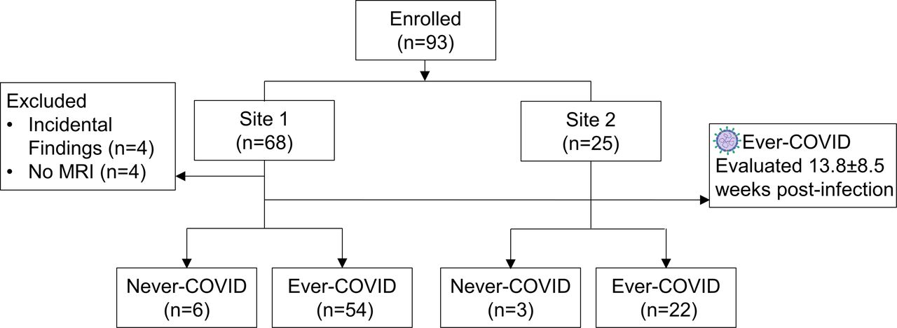
CONSORT (Consolidated Standards of Reporting Trials) diagram. Of the 93 participants enrolled, 68 participants were enrolled from Site 1 and 25 were enrolled from Site 2. Of the 68 participants enrolled from Site 1, 4 were excluded due to incidental findings, 4 were excluded because MRI was not acquired, 6 were never-COVID participants and 54 were ever-COVID participants. Of the 25 participants enrolled from Site 2, 3 were never-COVID participants and 22 were ever-COVID participants.
Table 1 provides a summary of baseline characteristics for 9 never-COVID and 76 ever-COVID participants including 23 (30%) ever-hospitalised and 53 (70%) never-hospitalised participants. There were significant differences for age (p<0.001) and sex (p=0.001) between participant subgroups. Of the 23 hospitalised participants, 3 required mechanical ventilation, 1 required non-invasive ventilation and 18 were provided supplemental oxygen during their hospital admission. Online supplemental table S1 provides baseline demographics by site and shows that the number of weeks between a positive COVID-19 test and the research visit (p=0.01), as well as SpO2 (p=0.01) were significantly different between the two sites. Online supplemental table S2 provides a by-participant list of current medications and online supplemental table S3 provides a summary of these medications.
Supplemental material
Demographic characteristics
Imaging and other endpoints
Figure 2 shows representative 129Xe ventilation MRI (cyan) co-registered with anatomical 1H MRI (greyscale) for three never-COVID and four ever-COVID participants, for which there is also representative coronal CT and segmented airway trees. For never-COVID volunteers, there was visually obvious homogeneous ventilation and VDP was <1% (P77 VDP=0.9%, P78 VDP=0.7%, P79 VDP=0.6%). For ever-COVID participants, there was visually obvious ventilation heterogeneity or defects and yellow arrows point to regions of interest with ventilation or CT abnormalities. In participant P26, there was a large ventilation defect in the right upper lobe that spatially corresponded to a radiodense region visible on CT. In participant P53, there were ventilation defects in the periphery of the upper lobes. In participants P14 and P02, there were large ventilation defects in the lung periphery. Online supplemental table S4 shows CT diagnostic findings, for a subset of participants with CT reports completed by a radiologist, including ground-glass opacities (20/50, 40%), atelectasis (14/50, 28%), nodules (13/50, 26%) and bronchiectasis (11/50, 22%).
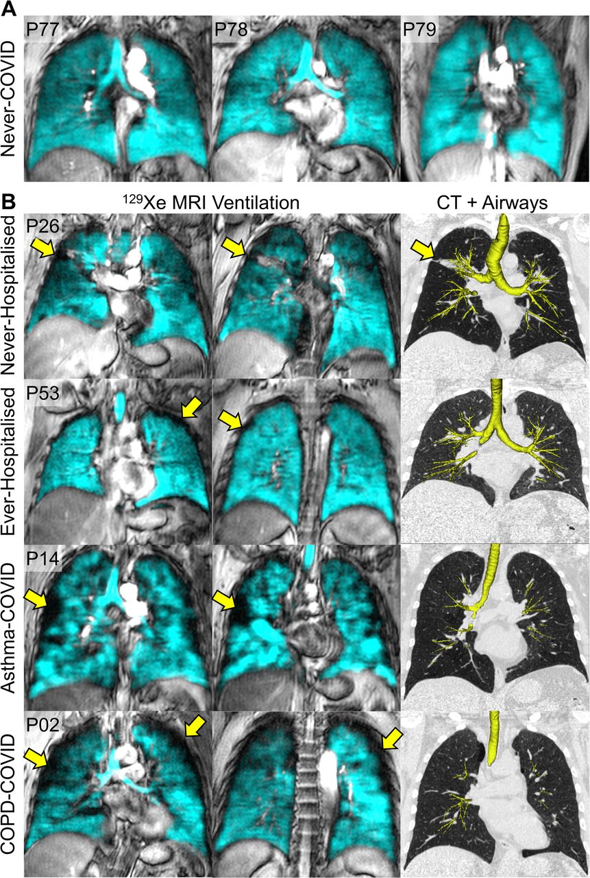
Representative 129Xe MRI Ventilation and CT airways. (A) Centre coronal 129Xe MRI slice (cyan) co-registered with 1H MRI thoracic cavity (greyscale) in three never-COVID participants (P77: female, VDP=0.9%; P78: female, VDP=0.7%; P79: female, VDP=0.6%). (B) Two coronal 129Xe MRI slices (cyan) co-registered with 1H MRI thoracic cavity (greyscale) and CT with segmented airway tree in four ever-COVID participants. Yellow arrows point to MRI or CT abnormalities. P26 was a male with no prior respiratory illness and FEV1=95%, LCI=8.9, SGRQ=36, mMRC=1, VDP=4.2% and TAC=475. P53 was a male with no prior respiratory illness and FEV1=87%, LCI=8.2, SGRQ=51, mMRC=1, VDP=2.8% and TAC=301. P14 was a female with asthma and never-hospitalised, FEV1=71%, SGRQ=37, mMRC=0, VDP=17.7% and TAC=289. P02 was a male with COPD and ever-hospitalised, FEV1=96%, LCI=8.0, SGRQ=14, mMRC=0, VDP=8.0% and TAC=169. COPD, chronic obstructive pulmonary disease; FEV1, forced expiratory volume in 1 s; LCI, lung clearance index; mMRC, modified Medical Research Council; SGRQ, St. George’s Respiratory Questionnaire; TAC, total airway count; VDP, ventilation defect per cent.
These qualitative findings are reflected in the quantitative findings shown in table 2 which summarises pulmonary function test, imaging and questionnaire data. Online supplemental table S5 shows all pulmonary function test, imaging and questionnaire data by site, while online supplemental table S6 and figure S1 show these findings by previously diagnosed asthma and COPD.
Pulmonary function, questionnaire and imaging measurements
As shown in table 2, there were significant differences among subgroups for FEV1 (p=0.005), FVC (p=0.004), LCI airway opening (LCIao) (p=0.04), DLCO (p=0.009), 6MWD (p=0.005), post-exertional SpO2 (p=0.03) and 129Xe MRI VDP (p=0.03). In addition, mean VDP (≥4.3%),43 SGRQ (≥6)45 and LCI/LCIao (≥7.5)46 were abnormal in ever-COVID as well as the ever-hospitalised and never-hospitalised participant subgroups.
Box and whisker plots in figure 3 show that 129Xe MRI VDP was significantly greater in ever-COVID as compared with never-COVID (p<0.001), and in ever-hospitalised as compared with never-COVID (p=0.01) participants and was not significantly different between never-hospitalised as compared with never-COVID participants (p=0.2). 129Xe MRI VDP was also significantly worse in ever-hospitalised versus never-hospitalised COVID-19 participants (p=0.048). There were also significant differences for DLCO (p=0.009) and 6MWD (p=0.005) between ever-hospitalised and never-hospitalised COVID-19 participants. Online supplemental figure S1 shows that 129Xe MRI VDP was significantly worse in participants with prior COPD as compared with COVID-19 patients with prior asthma (p=0.002) and those with no prior respiratory diagnosis (p<0.001). The 6MWD was also significantly worse (p=0.02) in participants with prior COPD as compared with those with no prior respiratory diagnosis. Participants were dichotomised by VDP (normal VDP <4.3%, total n=49, hospitalised n=15; abnormal VDP ≥4.3%, total n=27, hospitalised n=8). As shown in online supplemental figure S2, participants with abnormal VDP also had significantly worse 6MWD (p=0.003) and post-exertional SpO2 (p=0.03).
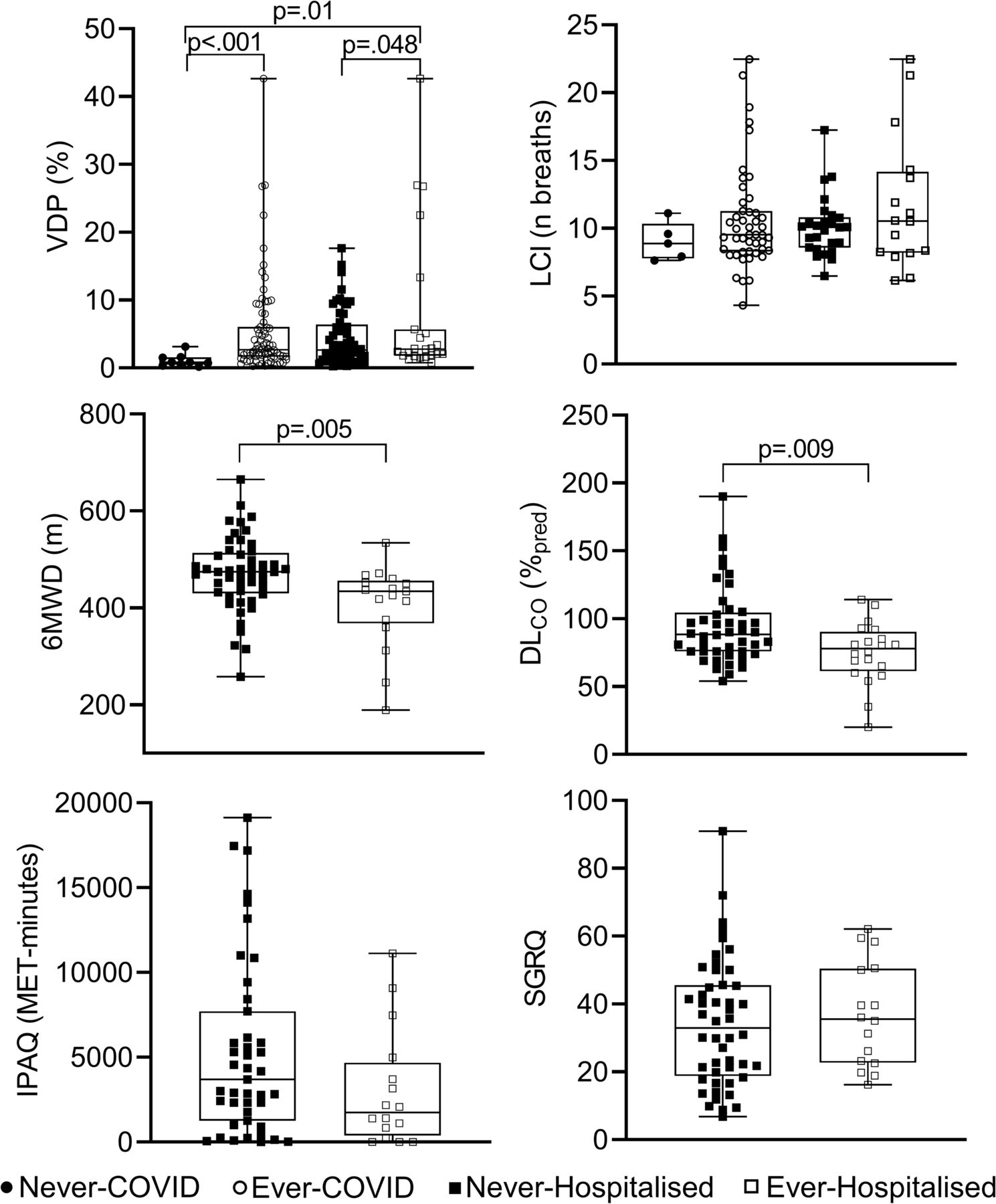
129Xe MRI VDP, pulmonary function, exercise capacity and quality-of-life VDP significantly different for ever-COVID versus never-COVID (p<0.001), and ever-hospitalised versus never-hospitalised (p=0.048) and never-COVID (p=0.01). 6MWD significantly different for ever-hospitalised versus never-hospitalised (p=0.005). DLCO significantly different for ever-hospitalised versus never-hospitalised (p=0.009). LCI, IPAQ and SGRQ not significantly different. DLCO, diffusing capacity of the lung for carbon-monoxide; IPAQ, International Physical Activity Questionnaire; LCI, lung clearance index; SGRQ, St. George’s Respiratory Questionnaire; VDP, ventilation defect per cent; 6MWD, 6-minute walk distance; %pred, per cent of predicted value.
Relationships
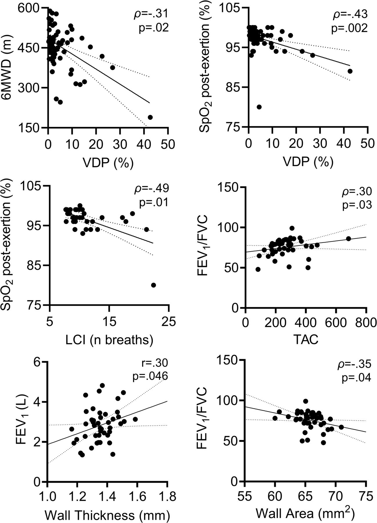
Figure 4 shows linear relationships for imaging and pulmonary function measurements. For completeness and transparency, online supplemental table S7 provides the Pearson or Spearman correlations for all parameters measured. As shown in figure 4, there were significant relationships for VDP with 6MWD (ρ=–0.31; p=0.02) and post-exertional SpO2 (ρ=–0.43; p=0.002) and for post-exertional SpO2 with LCI (ρ=–0.49; p=0.01). CT measurement relationships included TAC with FEV1/FVC (ρ=0.30, p=0.03), CT wall thickness with FEV1 (r=0.30, p=0.046) and CT wall area with FEV1/FVC (ρ=–0.35, p=0.04).
Relationships for MRI and CT Measurements. Linear correlation for VDP and 6MWD (ρ=−0.31, p=0.02). Linear correlation for VDP and SpO2 post-exertion (ρ=−0.43, p=0.002). Linear correlation for LCI and SpO2 post-exertion (ρ=−0.49, p=0.01). Linear correlation for TAC and FEV1/FVC (ρ=0.30; p=0.03). Linear correlation for wall thickness and FEV1 (r=0.30; p=0.046). Linear correlation for wall area and FEV1/FVC (ρ=−0.35; p=0.04). FEV1, forced expiratory volume in 1 s; FVC, forced vital capacity; LCI, lung clearance index; TAC, total airway count; VDP, ventilation defect per cent; 6MWD, 6-minute walk distance; ρ, Spearman correlation; r, Pearson correlation; p, Holm-Bonferroni corrected p value.
Discussion
To our knowledge, this is the first multicentre study that explores ventilation heterogeneity in ever-hospitalised and never-hospitalised post-COVID-19 participants with persistent and serious symptoms necessitating clinical follow-up. In 76 participants, we observed: (1) significantly different (worse) MRI VDP in ever-COVID as compared with never-COVID, and in never-hospitalised as compared with ever-hospitalised COVID-19 participants, who also experienced significantly different (worse) DLCO and 6MWD, (2) significant relationships between CT airway and spirometry measurements and (3) significant relationships between post-exertional SpO2 with two measurements of ventilation heterogeneity, MRI VDP and LCI, and between MRI VDP and the 6MWD.
First, we observed that MRI VDP was significantly different in ever-COVID as compared with never-COVID participants and mean VDP in ever-COVID patients was consistent with previously reported values20 in asymptomatic ever-hospitalised patients, post discharge. The finding of significantly different MRI VDP in ever-hospitalised as compared with never-hospitalised participants was novel as was the coincident finding of significantly different 6MWD and post-exertional SpO2 in these participants. Previous studies have observed worse 6MWD in severe versus mild–moderate post-COVID patients,47–49 however, the classification for mild, moderate or severe infection was not based on prior hospitalisation and not all participants experienced ongoing COVID-19 symptoms. When dichotomised by VDP, 6MWD and post-exertional SpO2 were also significantly different between groups. The link between exercise limitation, post-exertional oxygen saturation and ventilation heterogeneity in PACS is novel and points to a mechanistic relationship between persistent symptoms and airway pathologies and/or occlusion/obliteration.
Underscoring these central results was the finding of significant relationships for post-6MWD SpO2 with MRI VDP and with LCI—both are measurements of ventilation heterogeneity that are thought to reflect airway inhalational (MRI) and exhalational (LCI) function. This is the first supportive evidence of a potential link between exercise intolerance following COVID-19 and objective (LCI and MRI VDP) and direct (MRI VDP) measurements of abnormal airway function. This evidence was also supported by the significant associations between CT findings including total airway count (reflecting airway narrowing or obliteration/occlusion), airway lumen area (narrowing or occlusion) and airway wall thickness with spirometry measurements of airflow obstruction. Recent studies have provided evidence of small airways disease in post-COVID-19 patients using the full-scale airway network flow model50 or CT air-trapping functional small airways disease.51 We did not acquire inspiratory–expiratory CT and thus could not quantify CT air-trapping, although the ratio of residual volume to total lung capacity (RV/TLC) was measured in 38 of our 76 participants and of these, 14 (37%) reported RV/TLC > upper limit of normal.52 In addition, the median RV/TLC value in these 38 participants was 0.41±0.17, suggesting air-trapping, and this value was greater than the evaluation group reported by Cho et al (median RV/TLC=0.30).51 Taken together, these findings support the use of airways disease treatments, including inhaled combination corticosteroid–long-acting beta agonist, in post-COVID patients with persistent symptoms.
We acknowledge a number of limitations in this convenience-sample study, including the relatively small sample size. We should note that Site 1 participants were monitored over time by a long-COVID clinic designed to follow and treat long-term symptoms, including dyspnoea, and this clinic referred participants to consider enrolling in the study. Hence, there is a potential for bias towards a greater number and intensity of unexplained symptoms for participants enrolled. In addition, for most of the never-hospitalised participants enrolled, there was no prior clinical history of respiratory disease and thus first-time chest imaging and pulmonary function test results were reported. Therefore, we had to untangle any previous clinical history from the impact of COVID-19 on symptoms. Furthermore, pulmonary function tests, as well as CT and MR imaging measurements, prior to COVID-19 infection were not available, and thus, the impact of COVID-19 on existing lung function is still not completely clear. For a relatively large number of ever-COVID participants, chest CT was either declined (n=18) or not quantitatively evaluable (n=13) which also limits the generalisability of the CT results. It is also important to acknowledge that this was a multicentre study, with participants enrolled from two relatively diverse regional healthcare systems embedded within a population of 14.7 million people served by a single universal and comprehensive healthcare insurance plan. Site 1 enrolment derived from a quaternary care academic health centre serving a mainly rural population and Site 2 enrolment derived from another quaternary care academic health centre, located about 100 km away from Site 1 with a mainly urban population, hence our findings may not be generalisable to other healthcare jurisdictions. Moreover, we also recognise that the convenience study group we evaluated included a relatively large number of people with previously diagnosed asthma (n=21) and COPD (n=6), which is a greater proportion of patients with comorbid obstructive lung disease than previous reports.2–5 19 20 We expect that the presence of previous obstructive lung disease likely has complex interactions with post-COVID pulmonary measurements including MRI VDP. Importantly, there were no significant differences for SGRQ and IPAQ scores between participants with and without a previous diagnosis of obstructive lung disease, which underscores the severity of illness in PACS. Finally, as previously reported,53 older people are at greater risk for poor COVID-19 outcomes and there is also a complex relationship between age and MRI VDP,54 so age must be considered as an important factor in our results. It should also be noted that the ever-COVID group was significantly older compared with never-COVID and hence this may have influenced VDP measurements in this group. However, MRI VDP was still greater in the ever-COVID group than predicted based on age alone.55
In conclusion, we explored a potential role for MRI and CT pulmonary pathologies to help explain persistent and life-altering symptoms including exercise limitation in post-COVID patients. While mean SGRQ score was not normal in the PACS participants studied here, spirometry, DLCO and CT measurements were normal or nearly normal. This provides some context to the growing body of evidence that shows MRI VDP provides a sensitive measurement of abnormal physiology that, in the PACS participants studied here, may contribute to exercise limitation.
In this study, participants were unable to achieve pre-COVID work and day-to-day life activities, so we asked the question, is there an airways component to PACS that can be measured using MRI VDP and does this relate to quality-of-life? Yes, this exploratory study did point to a relationship between the lung pathologies that resulted in abnormal MRI VDP (or ventilation heterogeneity) as well as exercise limitation and abnormal post-exertional SpO2. SGRQ and IPAQ scores also were highly abnormal and very similar among ever-COVID participants. The study results point to a possible airways disease explanation for the persistent symptoms experienced following COVID-19 infection, which may help improve and target treatment.
Data availability statement
Data are available upon reasonable request.
Ethics statements
Patient consent for publication
Ethics approval
This study involves human participants and was approved by HSREB #113224 and HiREB #12672. Participants gave informed consent to participate in the study before taking part.
References
Supplementary materials
Supplementary Data
This web only file has been produced by the BMJ Publishing Group from an electronic file supplied by the author(s) and has not been edited for content.
Footnotes
HKK and MJM are joint first authors.
HKK and MJM contributed equally.
Contributors HKK, MJM, AMM, CV, NR, NBK and SS were responsible for data acquisition. HKK and MJM were responsible for data analysis and for preparing the first draft of the manuscript. EAH and MA assisted with CT acquisition and evaluated the CT data. CV, TH, ID, JN and PN were responsible for recruiting study participants and providing clinical input and interpretation of the data. GES, MSA, AO and MK supported the study design development and interpretation of the data. GP was responsible for study design, data analysis and interpretation as well as being the guarantor of study data integrity. All authors had an opportunity to review and revise the manuscript and approved its final submitted version.
Funding This study was funded by the Ministry of Health and Long-Term Care Ontario. AMM is supported by a Natural Sciences and Engineering Council (Canada) doctoral scholarship. GP, MK and SS are supported by the Canada Research Chair Program. Other contributions: The authors acknowledge the support of the London Health Sciences Centre COVID-19 clinic and Middlesex London Health Unit (which provided all COVID-19 testing and reporting), along with the participants who consented to this 2-year longitudinal study. David Reese, Carol Awde, Shannon Faseruk and Julie Lecomte performed MRI, Angela Wilson, Chynna Huang and Melanie Kjarsgaard were responsible for pulmonary function testing.
Competing interests The authors have reported to BMJ Open Respiratory Research the following: TH has received grants from Fisher & Paykel, honoraria for speaking engagements from AstraZeneca, and has participated on advisory boards for Sanofi, AstraZeneca and Valeo, outside the submitted work. JN has received honoraria for speaking engagements from AstraZeneca, Horizon Therapeutics and Vertix, outside the submitted work. PN has received grants from AstraZeneca, Teva, Sanofi, Foresee and Cyclomedica and personal fees from AstraZeneca, Teva, Equillium, Arrowhead Pharma and GlaxoSmithKline, outside the submitted work. SS has received grants from Cyclomedica, personal fees from Arrowhead Pharma and honoraria for speaking engagements from AstraZeneca, Novartis and Polarean, outside the submitted work. GP has received grants from AstraZeneca and Novartis and honoraria for speaking engagements from AstraZeneca, outside the submitted work. GES, MSA, AO, MK, SS and GP received grants from the Ministry of Health and Long-Term Care Ontario related to this work. HKK, MJM, AMM, CV, NR, EAH, NBK, MA and ID have no conflicts to declare.
Patient and public involvement Patients and/or the public were involved in the design, or conduct, or reporting, or dissemination plans of this research. Refer to the Methods section for further details.
Provenance and peer review Not commissioned; externally peer reviewed.
Supplemental material This content has been supplied by the author(s). It has not been vetted by BMJ Publishing Group Limited (BMJ) and may not have been peer-reviewed. Any opinions or recommendations discussed are solely those of the author(s) and are not endorsed by BMJ. BMJ disclaims all liability and responsibility arising from any reliance placed on the content. Where the content includes any translated material, BMJ does not warrant the accuracy and reliability of the translations (including but not limited to local regulations, clinical guidelines, terminology, drug names and drug dosages), and is not responsible for any error and/or omissions arising from translation and adaptation or otherwise.