

Delayed Surgical Management of Acute Type A Aortic Dissection in a Patient with Recent COVID-19 Infection and Post-COVID-19 Bronchopneumonia—Case Report and Review of Literature
 , ,
, ,  and
and 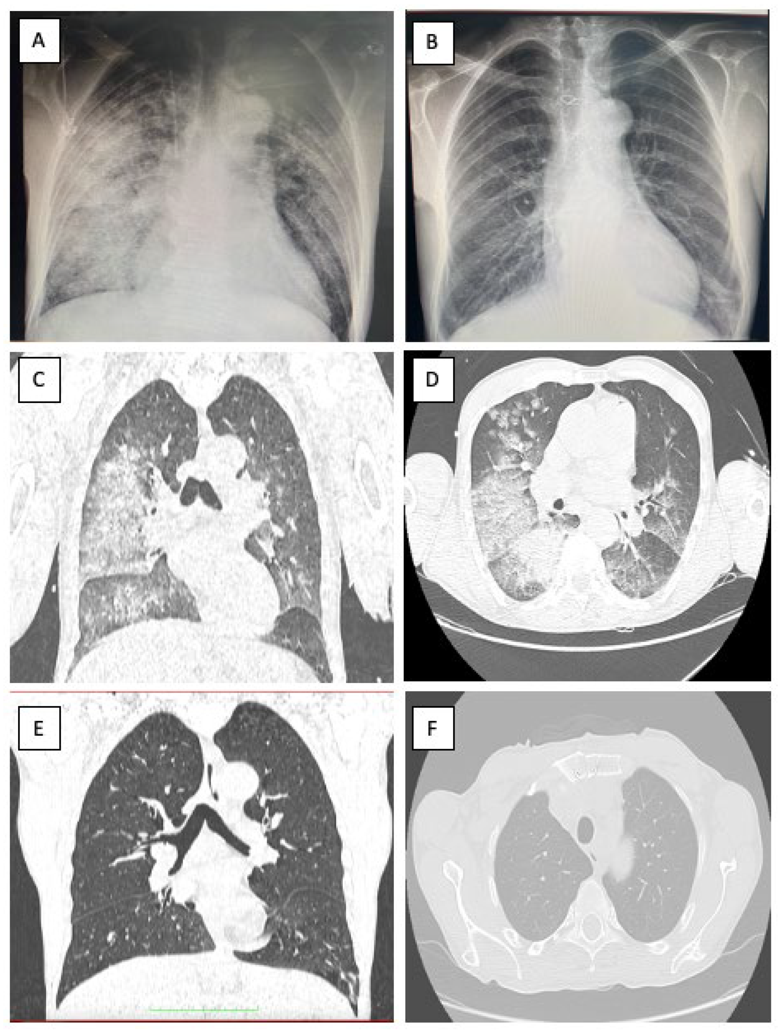
Abstract
:1. Introduction
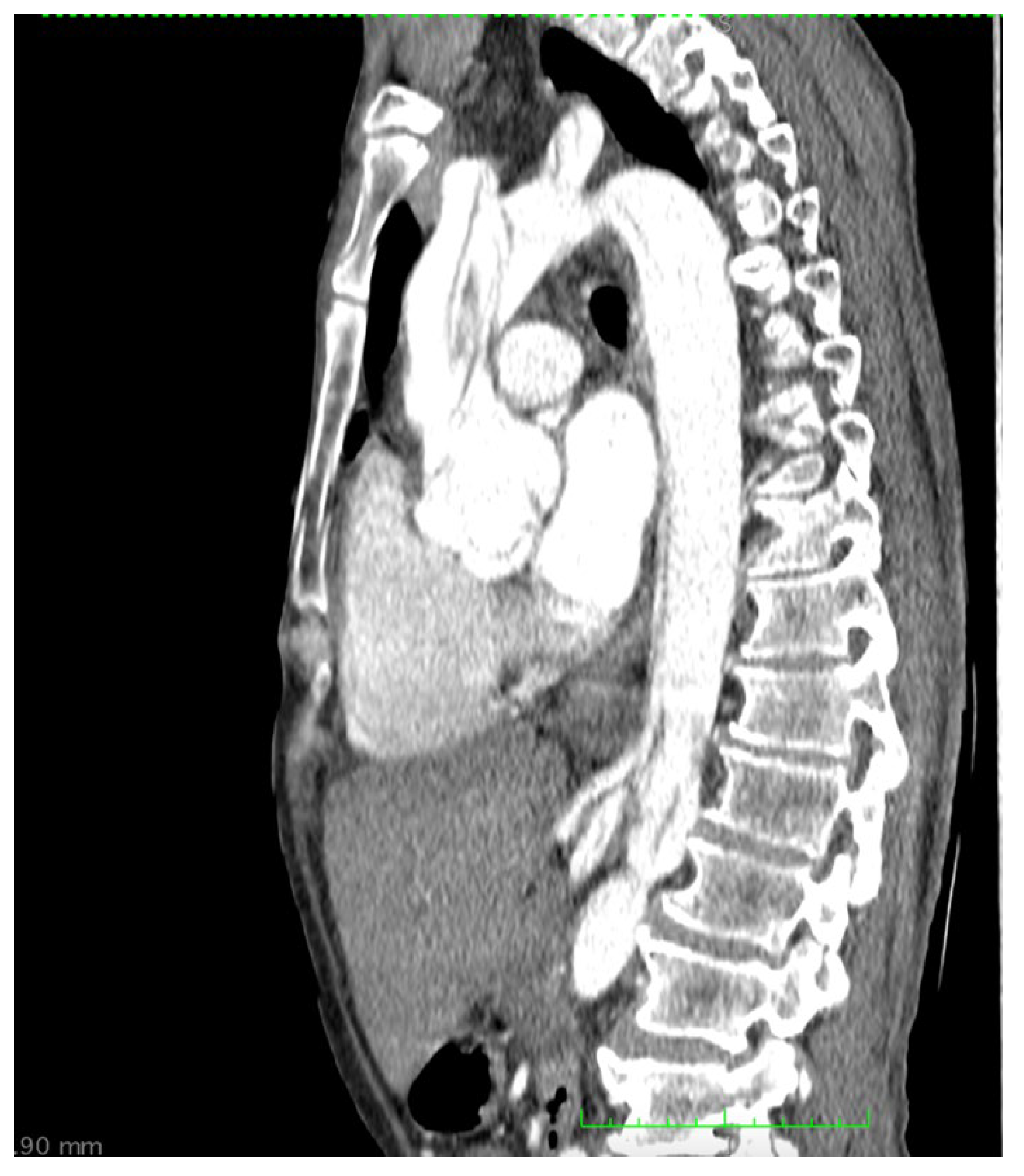
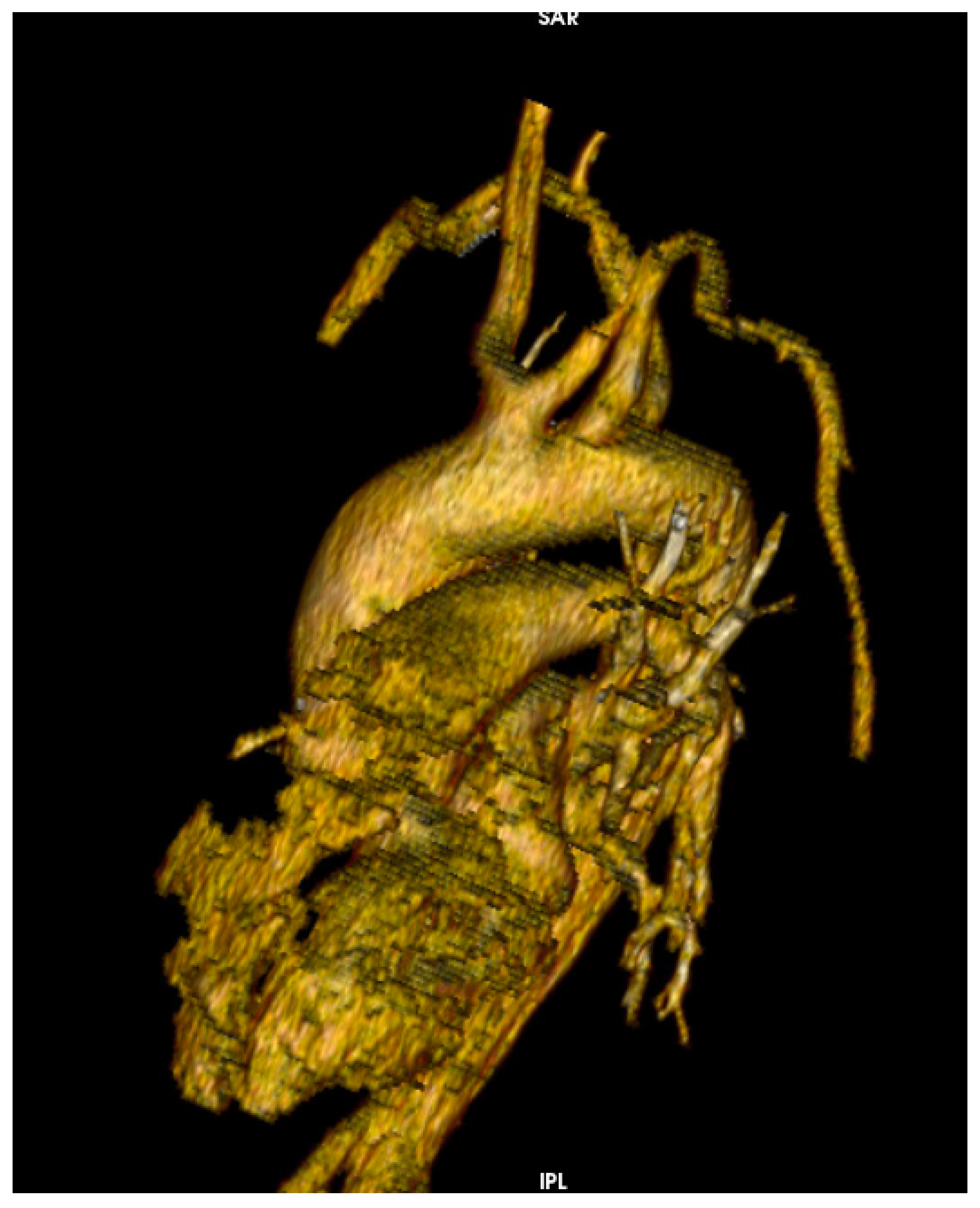
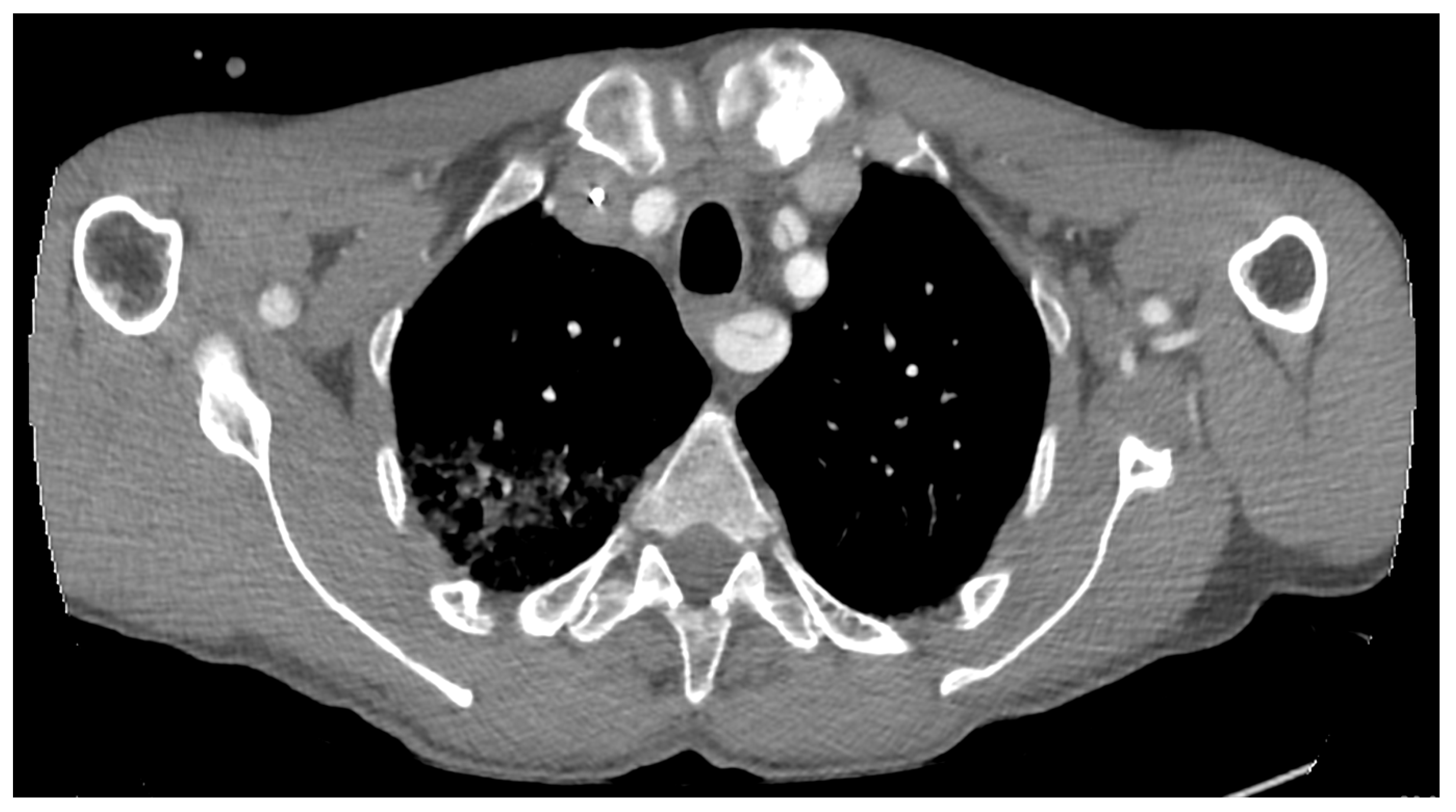
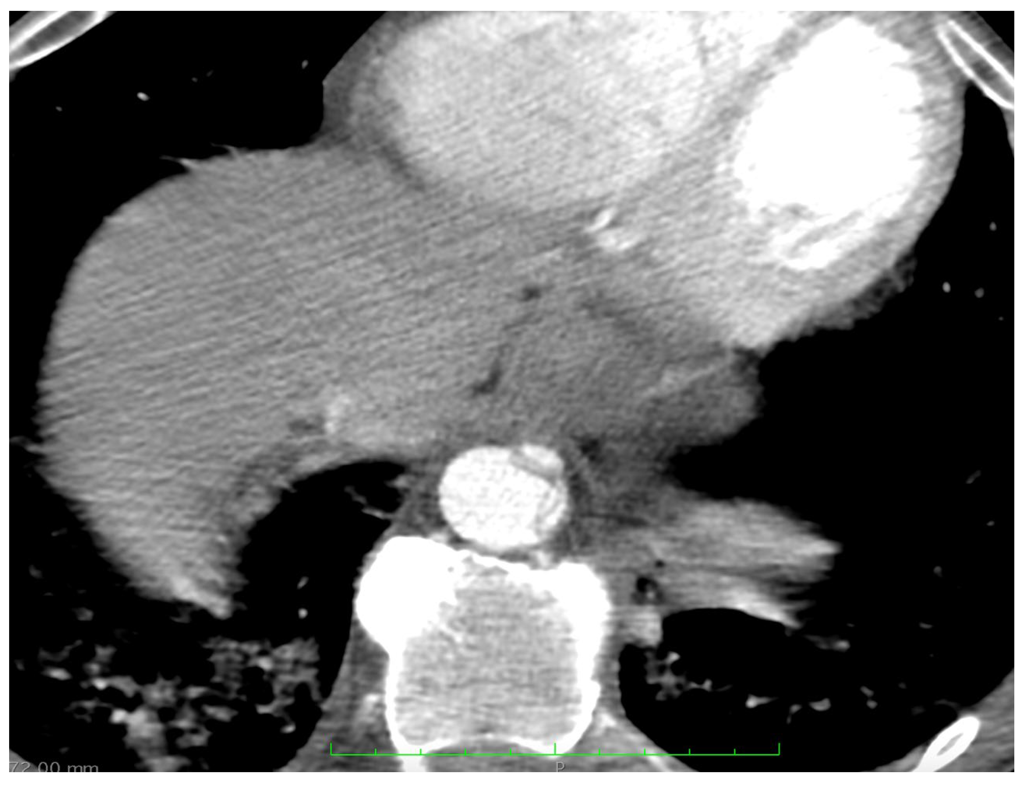
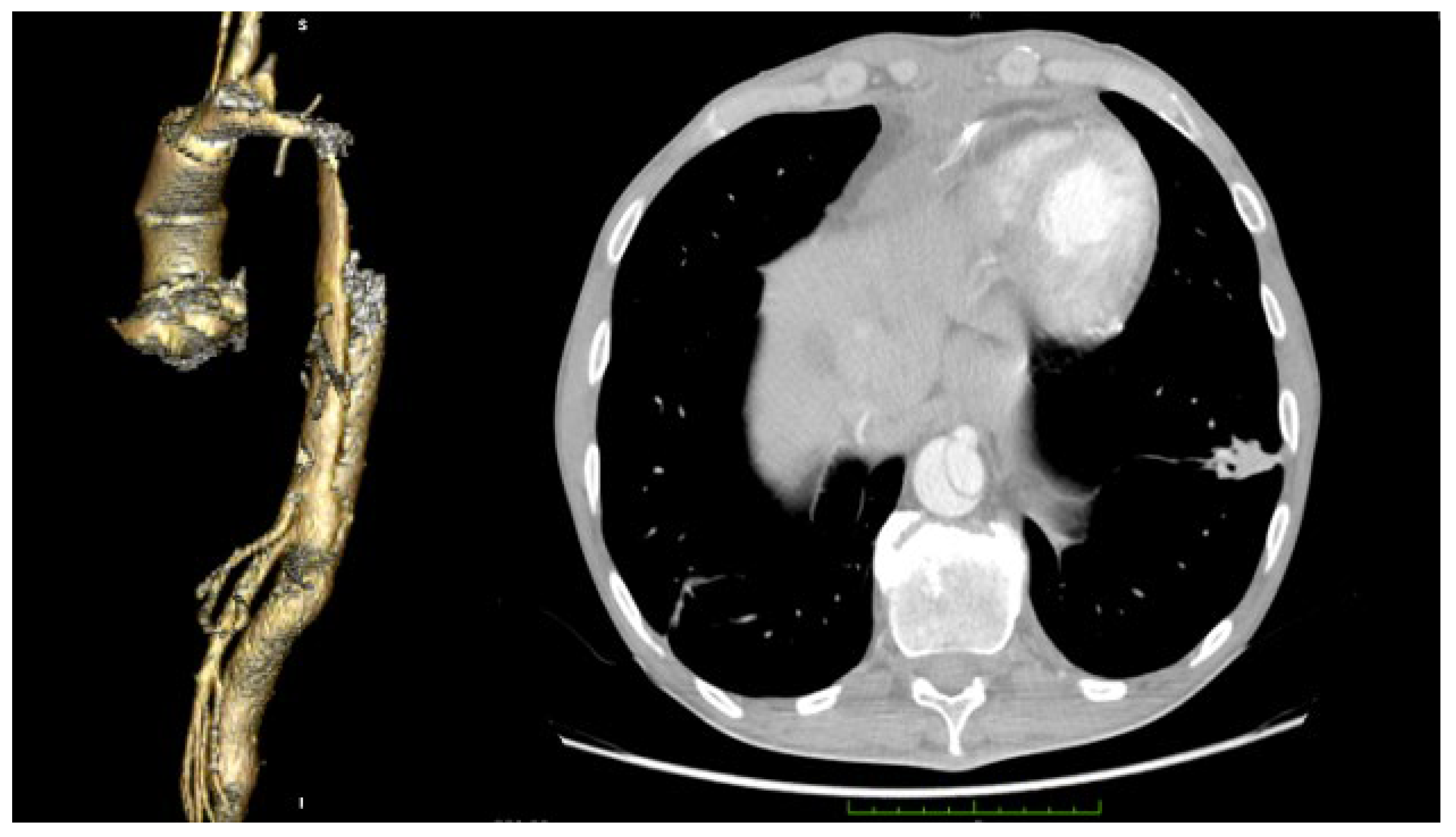
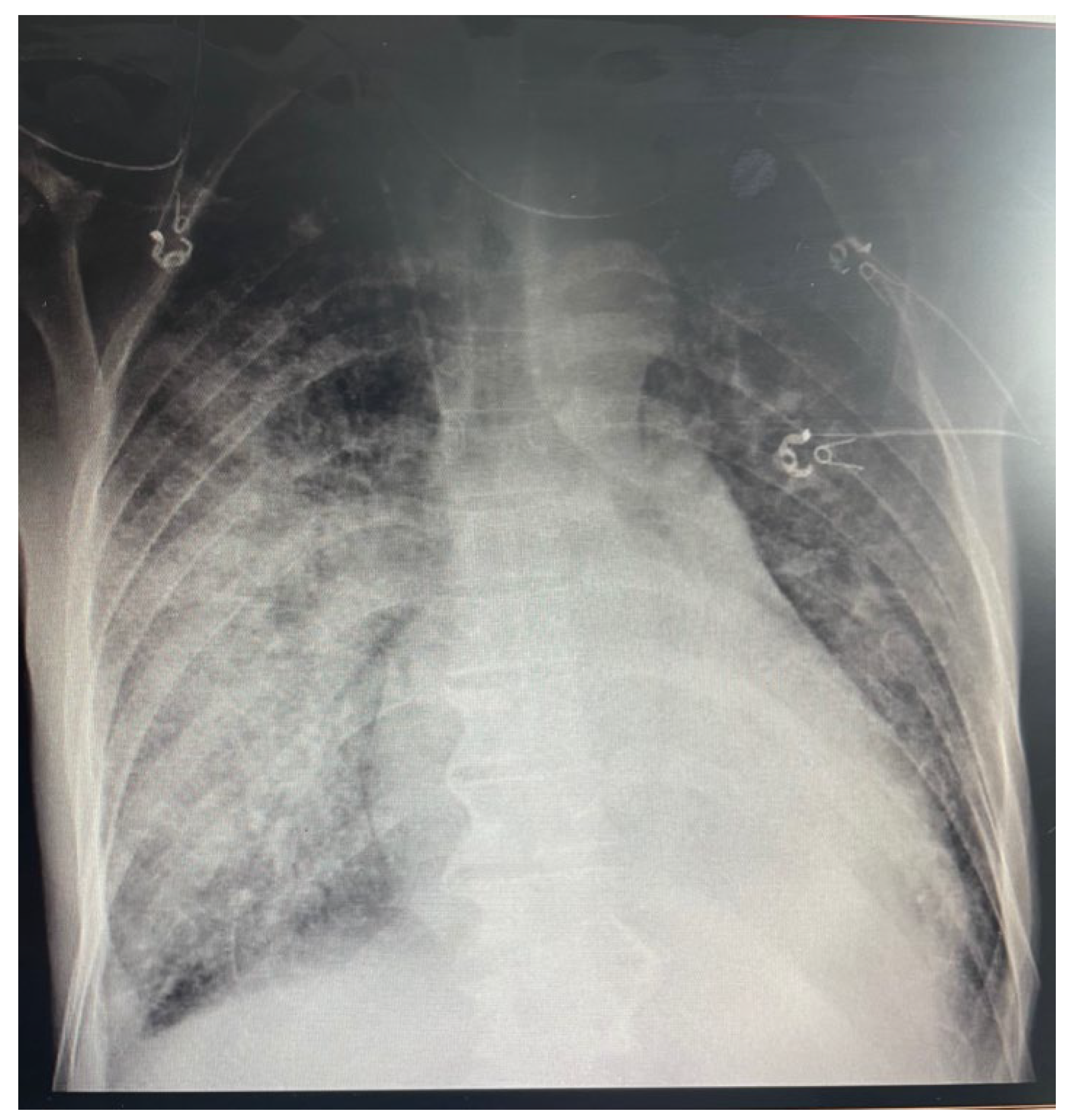
2. Case Report
3. Discussion
4. Conclusions
Author Contributions
Funding
Institutional Review Board Statement
Informed Consent Statement
Data Availability Statement
Conflicts of Interest
References
- Bedi, V.S.; Swain, P. and Yadav, A. Medical therapy versus TEVAR for uncomplicated type B aortic dissection. Indian J. Thorac. Cardiovasc. Surg. 2019, 35 (Suppl. S2), 174–178. [Google Scholar] [CrossRef] [PubMed]
- Rogers, R.K.; Reece, T.B.; Bonaca, M.P.; Hess, C.N. Acute Aortic Syndromes. Cardiol Clin. 2021, 39, 495–503. [Google Scholar] [CrossRef] [PubMed]
- Harky, A.; Singh, V.P.; Khan, D.; Sajid, M.M.; Kermali, M.; Othman, A. Factors Affecting Outcomes in Acute Type A Aortic Dissection: A Systematic Review. Heart Lung Circ. 2020, 29, 1668–1681. [Google Scholar] [CrossRef] [PubMed]
- Gupta, A.; Madhavan, M.V.; Sehgal, K.; Nair, N.; Mahajan, S.; Sehrawat, T.S.; Bikdeli, B.; Ahluwalia, N.; Ausiello, J.C.; Wan, E.Y.; et al. Extrapulmonary manifestations of COVID-19. Nat. Med. 2020, 26, 1017–1032. [Google Scholar] [CrossRef] [PubMed]
- Ghazavi, A.; Ganji, A.; Keshavarzian, N.; Rabiemajd, S.; Mosayebi, G. Cytokine profile and disease severity in patients with COVID-19. Cytokine 2021, 137, 155323. [Google Scholar] [CrossRef] [PubMed]
- Iskandar, Z.; Mordi, I.; Lang, C.C.; Huang, J.T.; Choy, A.-M. Biomarkers of aortopathy in Marfan syndrome. Cardiol. Rev. 2020, 28, 92–97. [Google Scholar] [CrossRef]
- Ponti, G.; Maccaferri, M.; Ruini, C.; Tomasi, A.; Ozben, T. Biomarkers associated with COVID-19 disease progression. Crit. Rev.Clin. Lab. Sci. 2020, 57, 389–399. [Google Scholar] [CrossRef]
- Arsene, A.L.; Dumitrescu, I.B.; Dragoi, C.M.; Udeanu, D.I.; Lupuliasa, D.; Jinga, V.; Drăgănescu, D.; Dinu-Pîrvu, C.E.; Burcea Dragomiroiu, G.T.A.; Blejan, I.E.; et al. A new era for the therapeutic management of the ongoing COVID-19 pandemic. Farmacia 2020, 68, 185–196. [Google Scholar] [CrossRef]
- Ramandi, A.; Akbarzadeh, M.A.; Khaheshi, I.; Khalilian, M.R. Aortic dissection and COVID-19; a comprehensive systematic review. Curr. Probl. Cardiol. 2022, 92, 101129. [Google Scholar] [CrossRef]
- Erbel, R.; Aboyans, V.; Boileau, C.; Bossone, E.; di Bartolomeo, R.; Eggebrecht, H.; Evangelista, A.; Falk, V.; Frank, H.; Gaemperli, O.; et al. 2014 ESC Guidelines on the diagnosis and treatment of aortic diseases: Document covering acute and chronic aortic diseases of the thoracic and abdominal aorta of the adult The Task Force for the Diagnosis and Treatment of Aortic Diseases of the European Society of Cardiology (ESC). Eur. Heart J. 2014, 35, 2873–2926. [Google Scholar] [CrossRef] [Green Version]
- Yang, B.; Norton, E.L.; Rosati, C.M.; Wu, X.; Kim, K.M.; Khaja, M.S.; Deeb, G.M.; Williams, D.M.; Patel, H.J. Managing patients with acute type A aortic dissection and mesenteric malperfusion syndrome: A 20-year experience. J. Thorac. Cardiovasc. Surg. 2019, 158, 675–687.e4. [Google Scholar] [CrossRef]
- Song, S.; Huh, U.; Lee, C.W.; Bae, M. Malperfusion in type A aortic dissection: One-staged hybrid treatment. Asian J. Surg. 2022, 45, 477–478. [Google Scholar] [CrossRef] [PubMed]
- Nienaber, C.A.; Powell, J.T. Management of acute aortic syndromes. Eur. Heart J. 2012, 33, 26–35b. [Google Scholar] [CrossRef] [PubMed]
- Mehta, R.H.; Suzuki, T.; Hagan, P.G.; Bossone, E.; Gilon, D.; Llovet, A.; Maroto, L.C.; Cooper, J.V.; Smith, D.E.; Armstrong, W.F.; et al. Predicting death in patients with acute type A aortic dissection. Circulation 2002, 105, 200–206. [Google Scholar] [CrossRef] [PubMed]
- Li, B.; Yang, J.; Zhao, F.; Zhi, L.; Wang, X.; Liu, L.; Bi, Z.; Zhao, Y. Prevalence and impact of cardiovascular metabolic diseases on COVID-19 in China. Clin. Res. Cardiol. 2020, 109, 531–538. [Google Scholar] [CrossRef]
- Wang, D.; Hu, B.; Hu, C.; Zhu, F.; Liu, X.; Zhang, J.; Wang, B.; Xiang, H.; Cheng, Z.; Xiong, Y.; et al. Clinical Characteristics of 138 Hospitalized Patients with 2019 Novel Coronavirus-Infected Pneumonia in Wuhan, China. JAMA 2020, 323, 1061–1069, Erratum in JAMA 2021, 325, 1113. [Google Scholar] [CrossRef] [PubMed]
- Oudit, G.Y.; Kassiri, Z.; Jiang, C.; Liu, P.P.; Poutanen, S.M.; Penninger, J.M.; Butany, J. SARS-coronavirus modulation of myocardial ACE2 expression and inflammation in patients with SARS. Eur. J. Clin. Investig. 2009, 39, 618–625. [Google Scholar] [CrossRef] [PubMed]
- Xiong, T.; Redwood, S.; Prendergast, B.; Chen, M. Coronaviruses and the cardiovascular system: Acute and long-term implications. Eur. Heart J. 2020, 41, 1798–1800. [Google Scholar] [CrossRef]
- Giusti, B.; Porciani, M.C.; Brunelli, T.; Evangelisti, L.; Fedi, S.; Gensini, G.F.; Abbate, R.; Sani, G.; Yacoub, M.; Pepe, G. Phenotypic variability of cardiovascular manifestations in Marfan Syndrome: Possible role of hyperhomocysteinemia and C677T MTHFR gene polymorphism. Eur. Heart J. 2003, 24, 2038–2045. [Google Scholar] [CrossRef]
- Shi, S.; Su, M.; Shen, G.; Hu, Y.; Yi, F.; Zeng, Z.; Zhu, P.; Yang, G.; Zhou, H.; Li, Q.; et al. Matrix metalloproteinase 3 as a valuable marker for patients with COVID-19. J. Med. Virol. 2021, 93, 528–532. [Google Scholar] [CrossRef]
- Petito, E.; Falcinelli, E.; Paliani, U.; Cesari, E.; Vaudo, G.; Sebastiano, M.; Cerotto, V.; Guglielmini, G.; Gori, F.; Malvestiti, M.; et al. Neutrophil more than platelet activation associates with thrombotic complications in COVID-19 patients. J. Infect. Dis. 2020, 223, 933–944. [Google Scholar] [CrossRef] [PubMed]
- Cardiothoracic Interdisciplinary Research Network and COVIDSurg Collaborative. Early outcomes and complications following cardiac surgery in patients testing positive for coronavirus disease 2019: An international cohort study. J. Thorac. Cardiovasc. Surg. 2021, 162, e355–e372. [Google Scholar] [CrossRef] [PubMed]
- Surg, J.C.; Niknam, J.; Rong, L.Q. Asymptomatic patients with coronavirus disease and cardiac surgery: When should you operate? J. Card. Surg. 2020, 35, 2486–2488. [Google Scholar]
- Hwang, D.; Zhan, Y. A Combination of Type A Aortic Dissection and COVID-19: Operative Mortality of 33%? Ann. Thorac. Surg. 2021, 111, 1734. [Google Scholar] [CrossRef] [PubMed]
- Stammers, A.H.; Mongero, L.B.; Tesdahl, E.A.; Patel, K.P.; Jacobs, J.P.; Firstenberg, M.S.; Petersen, C.; Barletti, S.; Gibbs, A. The assessment of patients undergoing cardiac surgery for COVID-19: Complications occurring during cardiopulmonary bypass. Perfusion 2022, 37, 350–358. [Google Scholar] [CrossRef] [PubMed]
- Patel, V.; Jimenez, E.; Cornwell, L.; Tran, T.; Paniagua, D.; Denktas, A.E.; Chou, A.; Hankins, S.J.; Bozkurt, B.; Rosengart, T.K. Cardiac surgery during the coronavirus disease 2019 pandemic: Perioperative considerations and triage recommendations. J. Am. Heart Assoc. 2020, 9, e017042. [Google Scholar] [CrossRef]
- Crawford, T.C.; Beaulieu, R.J.; Ehlert, B.A.; Ratchford, E.V.; Black, J.H. Malperfusion Syndromes in Aortic Dissection. Vasc. Med. 2016, 21, 264–273. [Google Scholar] [CrossRef]
- Deeb, G.M.; Williams, D.M.; Bolling, S.F.; Quint, L.E.; Monaghan, H.; Sievers, J.; Karavite, D.; Shea, M. Surgical delay for acute type A dissection with malperfusion. Ann. Thorac. Surg. 1997, 64, 1669–1675. [Google Scholar] [CrossRef]
- Jurcut, R.; Savu, O.; Bogdan, A.; Popescu, A.F.; Herlea, V.; Ginghina, H.M.C. Primary Cardiac Leiomyosarcoma When Valvular Disease Becomes a Vascular Surgical Emergency. Circulation 2010, 121, e415–e418. [Google Scholar] [CrossRef] [PubMed] [Green Version]


Publisher’s Note: MDPI stays neutral with regard to jurisdictional claims in published maps and institutional affiliations. |
© 2022 by the authors. Licensee MDPI, Basel, Switzerland. This article is an open access article distributed under the terms and conditions of the Creative Commons Attribution (CC BY) license (https://creativecommons.org/licenses/by/4.0/).
Share and Cite
Robu, M.; Marian, D.R.; Vasile, R.; Radulescu, B.; Stegaru, A.; Voica, C.; Nica, C.; Gheorghita, D.; Zaharia, O.; Iulian, A.; et al. Delayed Surgical Management of Acute Type A Aortic Dissection in a Patient with Recent COVID-19 Infection and Post-COVID-19 Bronchopneumonia—Case Report and Review of Literature. Medicina 2022, 58, 1357. https://doi.org/10.3390/medicina58101357
Robu M, Marian DR, Vasile R, Radulescu B, Stegaru A, Voica C, Nica C, Gheorghita D, Zaharia O, Iulian A, et al. Delayed Surgical Management of Acute Type A Aortic Dissection in a Patient with Recent COVID-19 Infection and Post-COVID-19 Bronchopneumonia—Case Report and Review of Literature. Medicina. 2022; 58(10):1357. https://doi.org/10.3390/medicina58101357
Chicago/Turabian StyleRobu, Mircea, Diana Romina Marian, Rasvan Vasile, Bogdan Radulescu, Alice Stegaru, Cristian Voica, Claudia Nica, Daniela Gheorghita, Ondin Zaharia, Antoniac Iulian, and et al. 2022. "Delayed Surgical Management of Acute Type A Aortic Dissection in a Patient with Recent COVID-19 Infection and Post-COVID-19 Bronchopneumonia—Case Report and Review of Literature" Medicina 58, no. 10: 1357. https://doi.org/10.3390/medicina58101357