Article

Computational Screening of Phenylamino-Phenoxy-Quinoline Derivatives against the Main Protease of SARS-CoV-2 Using Molecular Docking and the ONIOM Method

by
Suwicha Patnin
1,
Arthit Makarasen
1,*,
Pongsit Vijitphan
1,
Apisara Baicharoen
1,
Apinya Chaivisuthangkura
2,
Mayuso Kuno
2 and
Supanna Techasakul
1,*
1
Laboratory of Organic Synthesis, Department of Chemistry, Chulabhorn Research Institute, Laksi, Bangkok 10210, Thailand
2
Department of Chemistry, Faculty of Science, Srinakharinwirot University, Wattana, Bangkok 10110, Thailand
*
Authors to whom correspondence should be addressed.
Molecules 2022, 27(6), 1793; https://doi.org/10.3390/molecules27061793
Submission received: 16 February 2022 / Revised: 5 March 2022 / Accepted: 7 March 2022 / Published: 9 March 2022

Abstract

:
In the search for new anti-HIV-1 agents, two forms of phenylamino-phenoxy-quinoline derivatives have been synthesized, namely, 2-phenylamino-4-phenoxy-quinoline and 6-phenylamino-4-phenoxy-quinoline. In this study, the binding interactions of phenylamino-phenoxy-quinoline derivatives and six commercially available drugs (hydroxychloroquine, ritonavir, remdesivir, S-217622, N3, and PF-07321332) with severe acute respiratory syndrome coronavirus 2 (SARS-CoV-2) main protease (Mpro) were investigated using molecular docking and the ONIOM method. The molecular docking showed the hydrogen bonding and hydrophobic interactions of all the compounds in the pocket of SARS-CoV-2 main protease (Mpro), which plays an important role for the division and proliferation of the virus into the cell. The binding free energy values between the ligands and Mpro ranged from −7.06 to −10.61 kcal/mol. The molecular docking and ONIOM results suggested that 4-(2′,6′-dimethyl-4′-cyanophenoxy)-2-(4″-cyanophenyl)-aminoquinoline and 4-(4′-cyanophenoxy)-2-(4″-cyanophenyl)-aminoquinoline have low binding energy values and appropriate molecular properties; moreover, both compounds could bind to Mpro via hydrogen bonding and Pi-Pi stacking interactions with amino acid residues, namely, HIS41, GLU166, and GLN192. These amino acids are related to the proteolytic cleavage process of the catalytic triad mechanisms. Therefore, this study provides important information for further studies on synthetic quinoline derivatives as antiviral candidates in the treatment of SARS-CoV-2.

1. Introduction

Coronaviruses are enveloped, single-stranded RNA viruses that contain proteins and are covered with carbohydrates [1,2]. These pathogens can infect humans, mammals, and reptiles and may cause respiratory and digestive tract diseases in animals that can be easily spread to humans. In early 2020, a human disease caused by a new coronavirus type called severe acute respiratory syndrome coronavirus 2 (SARS-CoV-2) spread in many countries worldwide. One of the vital SARS-CoV-2 enzymes is main protease (3CLpro), also known as Mpro, which is a homodimer. Each dimer is composed of three domains, designated as cysteine protease domain I–III, which contain 306 amino acids. Domains I and II are antiparallel β-barrel structures, and domain III contains five α-helices in one antiparallel globular cluster connecting to domain II [3]. The substrate binding site of Mpro is specific to all coronaviruses and comprises four subsites. The enzyme active site of Mpro contains a catalytic dyad of histidine and cysteine residues that behave as general acid/general base nucleophiles. This active site is in the cleft between domain I and II. In addition, Mpro cleaves at least 11 specific sites within the polyprotein during the replication process and plays an essential role in transcription to a viral gene. There is a strong conservation of residues in the binding pocket of Mpro. Due to the essential functions of transcription and replication, Mpro is considered to be an important drug target for antiviral treatment [4,5,6,7].
Many medications have been used for SARS-CoV-2 treatment. However, appropriate drugs remain undiscovered; thus, indefinite drugs are being administered to patients, while suitable medications and vaccines are being explored [8,9]. Hydroxychloroquine is an antimalarial drug that inhibits heme detoxification when administered to parasitic patients. Hydroxychloroquine was selected as a treatment in the first stage of the epidemic infection of the SARS-CoV-2 virus [10]. Additionally, ritonavir, a protease inhibitor, is used as an antiretroviral drug for human immunodeficiency virus 1 (HIV-1), but with limitations due to side effects. Ritonavir is used for treating SARS-CoV-2 because of the similarities between the protease enzymes of SARS-CoV-2 and HIV-1 [11]. Recently, physicians have reported that remdesivir, a nucleoside analog drug, inhibited virus multiplication, and it has been applied against Ebola disease, MERS, and SARS [12,13]. To date, three significant drugs have been found that inhibit the function of the protease enzymes in SARS-CoV-2, namely, S-217622, N3, and PF-07321332. The oral administration of S-217622, which is a main protease inhibitor, is in a Phase III clinical trial for SARS-CoV-2 treatment. The S-217622 drug showed a preclinical pharmacokinetic profile that supported daily oral dosing as an oral therapeutic agent for treating SARS-CoV-2 [14]. N3 is also a potent inhibitor of main protease in coronaviruses, especially SARS-CoV-2. The drug N3 has exhibited potent Mpro inhibitor activity, with an EC50 value of ≥16.77 µM, for the treatment of animal infections [15,16]. However, N3 displays hepatotoxicity, which makes it carcinogenic for humans [17]. The drug PF-07321332 is an oral form developed from PF-07304814, a potent main protease inhibitor in vitro selective to human protease targets. The PF-07321332 structure consists of a nitrile warhead acting as a Michael acceptor. In addition, PF-07321332 displays potent antiviral activity against SARS-CoV-2, with an EC90 value of 0.181 µM [18]. PF-07321332 prevents SARS-CoV-2 replication by inhibiting primary protease, which cleaves long protein chains to the crucial parts and binds to a cysteine residue in the cysteine protease enzyme [19,20]. Nowadays, PF-07321332 is available and authorized by regulatory bodies throughout the world, such as the EMA (in the European Union) [21]. Therefore, appropriate medications against SARS-CoV-2 are under development.
In the search for novel HIV-1 non-nucleoside reverse transcriptase inhibitors (NNRTIs), phenylamino-phenoxy-quinoline derivatives have been synthesized from a combination of the pharmacophore templates of nevirapine (NVP), efavirenz (EFV), and rilpivirine (TMC278) [22,23]. These compounds can be divided into two groups, 4,6-disubstituted quinoline (14) and 2,4-disubstituted quinoline (58), and have been evaluated for their inhibitory effect on HIV-1 reverse transcriptase. A previous study indicated that 2-phenylamino-4-phenoxy-quinoline derivatives exhibited similar percentage inhibitory activity to that of NVP. The study found that those compounds might interact and bind in a similar area to NNRTIs within the pocket of HIV-1 RT. Moreover, 2-phenylamino-4-phenoxy-quinoline derivatives have shown high cytotoxic activity against some human cancer cells over normal cells and excellent binding interactions with transported proteins [24]. Because phenylamino-phenoxy-quinoline derivatives are nitrogen-containing compounds with nitrile side chains, similar to current available drugs for SARS-CoV-2, they are considered to be candidate antiviral drugs for SARS-CoV-2 treatment.
In this study, the binding interactions of presently used medications and phenylamino-phenoxy-quinoline derivatives (18) with SARS-CoV-2 main protease (Mpro) were investigated using molecular docking and the ONIOM method. The structures of the compounds are shown in Figure 1. Molecular docking and the ONIOM method were used to describe the binding positions and binding interactions among biomolecules, and the results were presented as binding free energy and interaction energy values. Molecular docking and the ONIOM method have been successfully applied in most pharmaceutical research and modern drug discoveries [25,26]. Binding interactions are useful and important for understanding the function of the binding and inhibition processes. Therefore, this study has great potential for evaluating drug development for SARS-CoV-2 treatment.

2. Results and Discussion

2.1. Pharmacokinetics Study

The pharmacokinetics of the compounds were calculated using SwissADME (Developed and maintained by the Molecular Modeling Group of the SIB, Swiss Institute of Bioinformatics, Lausanne, Switzerland), which predicts the molecular properties and pharmacokinetic activity of drug-likeness. Drug-likeness is applied in screening drug candidates and is evaluated by Lipinski’s rule of five. Lipinski’s rule of five describes the absorption or permeation oral activity of a drug for the initial screening of drug-likeness and states that the molecular weight of compounds must be less than 500 Da, lipophilicity (log P) must be less than 5, H-bond donors must be less than 5, and H-bond accepters must be less than 10. Furthermore, the total polar surface area (TPSA) should be less than 140 Å2, and the number of rotatable bonds should be less than 10 [27,28]. According to pharmacokinetic studies of compounds, only hydroxychloroquine, PF-07321332, and compounds (18) correspond with Lipinski’s rule of five, together with their number of rotatable bonds being less than 10 (except PF-07321332) and TPSA less than 140 Å2, as shown in Table 1. Therefore, hydroxychloroquine, PF-07321332, and phenylamino-phenoxy-quinoline derivatives (18) are in accordance with Lipinski’s rule of five and are appropriate candidate drugs against SARS-CoV-2 main protease.

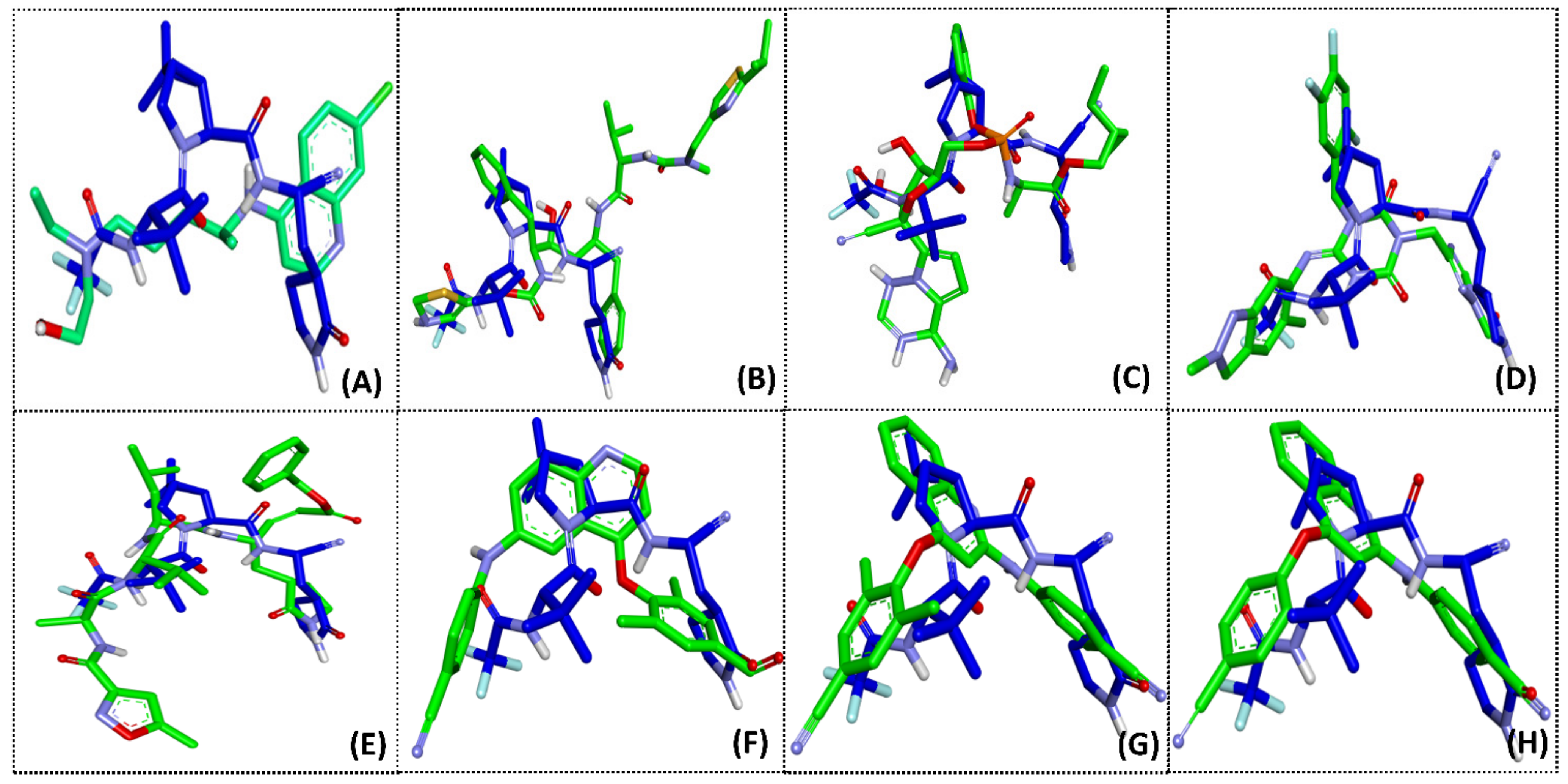
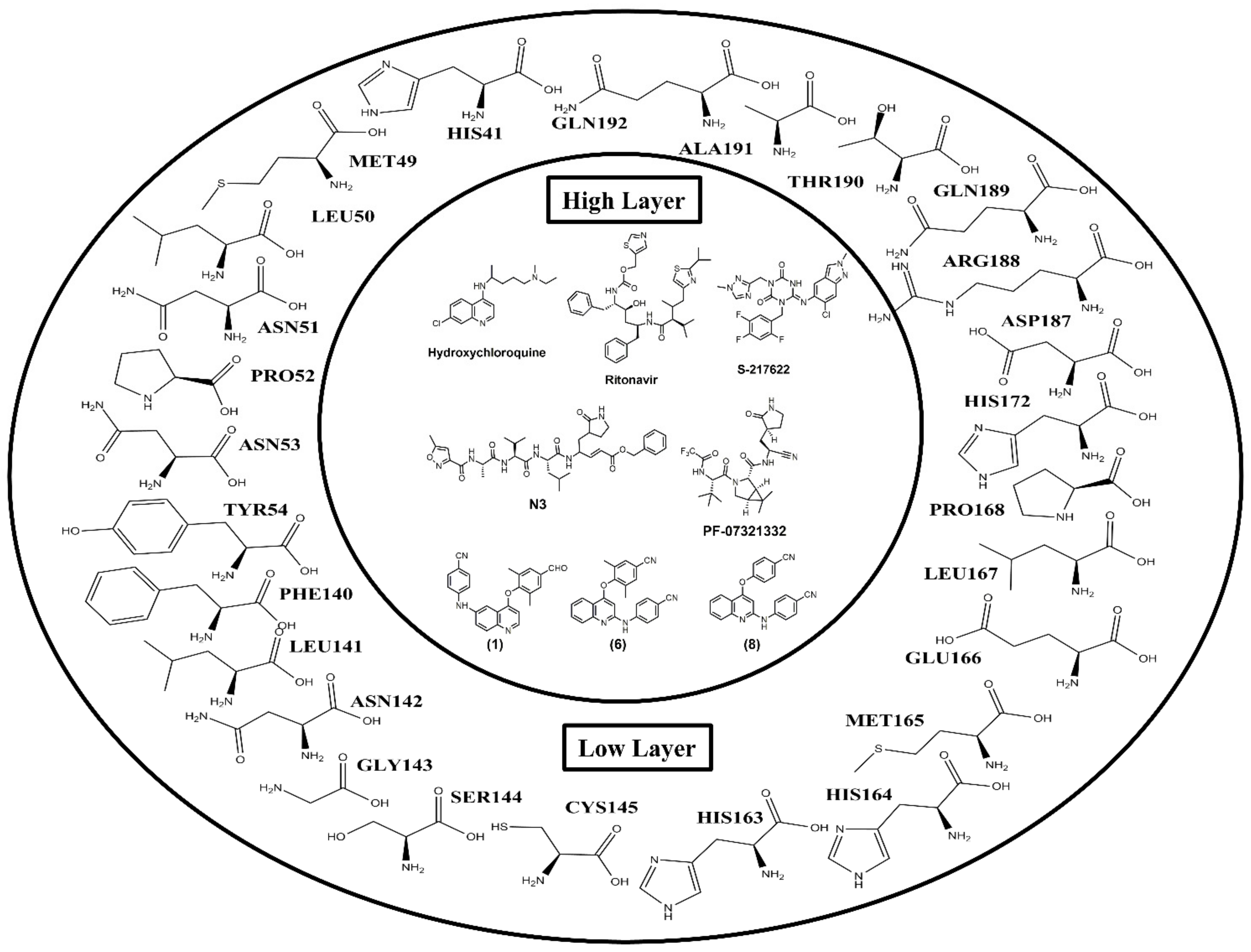
2.2. Molecular Docking

The binding interactions between the ligands in Mpro and the current medications, as well as the phenylamino-phenoxy-quinoline derivatives (compounds 18), were studied using molecular docking to understand the bonding mode of the compounds in Mpro and to provide information for drug design. The molecular docking results, as shown in Figure 2, indicate that all selected ligands bind to the hydrophobic cavity in the binding pocket of Mpro (Figure 2A). Moreover, the overlaying of each selected ligand in the binding pocket reveals that all compounds are bound in a similar region (Figure 2B). The binding sites of these ligands in Mpro were similar to the results of previous research, in which the active compounds against SARS-CoV-2 Mpro interacted with the amino acid residues, namely, HIS41, ASN142, GLY143, SER144, CYS145, GLU166, ASP187, THR190, and GLN192, in the substrate binding pocket of Mpro via hydrogen bonding and hydrophobic interactions [18,19,20].
The binding energy values between the selected ligands and Mpro ranged from −7.06 to −10.61 kcal/mol, as shown in Table 2. The three lowest binding energy values of −10.61, −10.12, and −10.02 kcal/mol were observed for PF-07321332, (4-(2′,6′-dimethyl-4′-formylphenoxy)-6-(4″-cyanophenyl)-aminoquinoline (1), and 4-(2′,6′-dimethyl-4′-cyanophenoxy)-2-(4′cyanophenyl)-aminoquinoline (6), respectively. PF-07321332 had the lowest binding energy value of −10.61 kcal/mol compared with other ligands, whereas hydroxychloroquine had the highest binding energy value of −7.08 kcal/mol. However, the results showed that phenylamino-phenoxy-quinoline 1, 6, and 8 exhibited lower binding energy than the current medications, except for PF-07321332. Ritonavir, S-217622, and N3, a protease inhibitor, displayed binding energy values of −8.56, −9.62, and −9.44 kcal/mol with Mpro, respectively. Remdesivir, which inhibited the RNA-dependent RNA polymerase (RdRp) of SARS-CoV-2, showed a binding energy value of −8.63 kcal/mol with Mpro. PF-07321332 demonstrated potent in vitro antiviral activity against SARS-CoV-2, as well as activity against other coronaviruses.
Hydroxychloroquine, ritonavir, remdesivir, S-217622, N3, PF-07321332, and compounds (1), (6), and (8) were selected to further study the overlay of the binding interaction in the Mpro binding pocket and to compare the conformation of optimized ligands. The results showed that PF-07321332 had the lowest binding energy values compared with the other drugs and compounds. Then, PF-07321332 was selected to study the overlay of optimized ligands in order to consider the appropriate position in the ligand-binding sites. The optimized conformation of the compounds, namely, hydroxychloroquine, ritonavir, remdesivir, S-217622, N3, (1), (6), and (8) compared with PF-07321332 in the Mpro binding pocket are displayed in Figure 3. The results implied that S-217622, N3, (1), (6), and (8) were arranged in a similar position to PF-07321332 with the appropriate area in the pocket of Mpro, except for hydroxychloroquine, ritonavir, and remdesivir (Figure 3A–C), since these compounds, which exhibited low binding energy value, contained heterocyclic skeletons with similar functional groups, such as phenyl and nitrile groups. The structure of (1), (6), and (8) consisted of the substituent at the 4,6- and 2,4- positions of the quinoline core structure, which differed from hydroxychloroquine in the substituents bound with Mpro. Studies in the literature have revealed that (1), (6), and (8) have shown the potential to inhibit HIV-1 RT in vitro without cytotoxicity against normal cell lines [20]. Therefore, during drug development, phenylamino-phenoxy-quinoline derivatives demonstrate potential as candidates for inhibiting Mpro in SARS-CoV-2.
The analysis of the binding interaction between ligands and Mpro found that ligands can bind to the binding pocket of Mpro via hydrogen bonding and the Pi-Pi stacking interaction, as shown in Table 2. The formed hydrogen bonds are of two types: strong and weak. A strong hydrogen bond is formed when a hydrogen atom binds to a high electronegativity atom and is called conventional hydrogen bonding. A weak hydrogen bond is formed when a hydrogen atom binds to elements such as sulfur (S), carbon (C), or aromatic rings which serve as electron donors and, thus, are called sulfur-hydrogen bonding, carbon-hydrogen bonding, or Pi-donor-hydrogen bonding, respectively [29,30].
The molecular docking results of the binding interactions of six commercially available drugs and compounds (1), (6), and (8) with Mpro in the binding pocket surrounding a radius of 5.0 Å from Mpro indicated that all of these compounds bound to the hydrophobic cavity of Mpro, as shown in Table 3. The 2D diagrams display the types of contacts between 14 compounds and Mpro, as shown in Figures S1–S4 (see also Supplementary Materials). Phenylamino-phenoxy-quinoline (1), (6), and (8) bound to the amino acid residues CYS145, HIS164, GLN189, and GLN192 via conventional hydrogen bonding, to GLU166 via Pi-donor hydrogen bonding, and to HIS41 via Pi-Pi stacking interaction in the active site of Mpro. The commercially available drugs bound to the amino acid residues HIS41, PHE140, LEU141, ASN142, GLY143, SER144, CYS145, HIS164, GLU166, LEU167, GLN189, THR190, and GLN192 via conventional hydrogen bonding, to MET49, PRO52, PHE140, MET165, and GLN189 via carbon hydrogen bonding, and to HIS41 via Pi-Pi stacking interaction in the binding pocket of Mpro.
Studies in the literature indicate that the amino acid residues HIS41, CYS145, and ASP187 are incorporated in the catalytic triad found in the active cavity on the Mpro surface and are involved in proteolysis. HIS41 and CYS145 may inhibit the catalytic triad mechanism of the proteolytic cleavage process of the Mpro, thus preventing the virus from replicating and reducing intracellular infection [31,32,33]. Moreover, the amino acid residue GLU166 plays an essential role in connecting the substrate binding site with the dimer structure of SARS-CoV-2 Mpro [34,35,36]. According to these results, we conclude that 4-(4′-cyanophenoxy)-2-(4″-cyanophenyl)-aminoquinoline (8) may have potential activity at a level similar to that of PF-07321332, because both compounds bind to Mpro via three amino acid residues, namely, HIS41, CYS145, and GLU166, at levels higher than other analyzed compounds.

2.3. ONIOM Study

In this study, ONIOM calculations were used to investigate the interaction mechanism of ligands with Mpro to obtain important information for developing new SARS-CoV-2 inhibitors. The system was divided into two layers: the high layer was treated using the B3LYP/6-31G(d,p) method, and the low layer was treated using the PM6 method (Figure 4). The interaction energy between the ligands and individual residues was calculated at the B3LYP/6-31G(d,p) level by using geometry from the X-ray structure. The interaction energy (Eint) between each ligand and the amino acid residue was calculated by using Equation (1). ONIOM calculations were applied to confirm the results of the binding interaction between each ligand, namely, hydroxychloroquine, ritonavir, S-217622, N3, PF-07321332, 1, 6, and 8, and the individual amino acid residues in Mpro, which demonstrated each of the optimal geometries from the results of molecular docking. The small model system consisted of 26 amino acid residues within the binding pocket surrounding Mpro in the radius of 5.0 Å, namely, HIS41, MET49, LEU50, ASN51, PRO52, ASN53, TYR54, PHE140, LEU141, ASN142, GLY143, SER144, CYS145, HIS163, HIS164, MET165, GLU166, LEU167, PRO168, HIS172, ASP187, ARG188, GLN189, THR190, ALA191, and GLN192.
The ONIOM calculations found that all compounds bound to the substrate binding site of Mpro with the interaction energy values, as shown in Table 4. The three lowest total interaction energy values were found in the case of N3, PF-07321332 and S-217622, with the values of −44.73, −49.51 and −40.25 kcal/mol, respectively. These results showed that phenylamino-phenoxy-quinoline (1), (6), and (8) exhibited lower interaction energy than hydroxychloroquine and ritonavir. Considering phenylamino-phenoxy-quinoline derivatives, compound 1 (4,6-disubstituted quinoline) and compounds 6 and 8 (2,4-disubstituted quinoline), (6) and (8) demonstrated lower interaction energy than (1). Consequently, the 2,4-disubstituted quinoline derivatives interacted more with MPro than the 4,6-disubstituted quinoline derivative.
The interaction energy values between the amino acid residues, namely, HIS41, GLU166, GLN189, and GLN192 and selected ligands showed the most significant contributions because of their lowest energy. GLU166 displayed the lowest interaction energy values with the ligands compared with other amino acid residues on Mpro, from −9.51 to −24.99 kcal/mol, indicating its important role in ligand interaction in the binding pocket of Mpro. In addition, the crucial interactions that occurred between ligands and amino acid residues such as MET49, PHE140, GLY143, SER144, CYS145, and HIS164 presented interaction energy from −0.02 to −5.02 kcal/mol. As described in the Molecular Docking Section 2.2, the amino acids HIS41, CYS145, and GLU166 play important roles in the replication and infection of the SARS-CoV-2 virus [32,33,34]. Interestingly, (1), (6), (8), and PF-07321332 interacted with Mpro using their link with amino acid residues namely, HIS41, CYS145, HIS164, GLU166, GLN189, and GLN192. The results corresponded with the molecular docking and suggest that hydrogen bonding and the Pi-Pi stacking interaction have the most substantial contributions to the binding interaction of these amino acid residues in the binding site of Mpro.
PF-07321332 and (8) showed efficient binding interactions with Mpro; bound with three amino acid residues, HIS41, CYS145, and GLU166; and exhibited lower interaction energy than (1) and (6). Meanwhile, (1) and (6) only interacted with HIS41 and GLU166. These results suggest that the binding activity of (8) is similar to that of PF-07321332. The results demonstrate that 4-(4′-cyanophenoxy)-2-(4″-cyanophenyl)-aminoquinoline (8) can be exploited to identify drug candidate compounds for the development of new anti-SARS-CoV-2 main protease agents.

3. Materials and Methods

3.1. Pharmacokinetics Study

Pharmacokinetics are used to define the absorption, distribution, metabolism, and excretion (ADME) parameters which explain the various characteristics of drugs in the body. In this study, SwissADME (Developed and maintained by the Molecular Modeling Group of the SIB, Swiss Institute of Bioinformatics, Lausanne, Switzerland) was applied to analyze the pharmacokinetic parameter of ligands [37]. Drug-likeness and molecular property prediction were screened depending on Lipinski’s rule of five [25,26].

3.2. Ligand and Protein Structure Preparation

Six commercially available drugs (hydroxychloroquine, ritonavir, remdesivir, S217622, N3, and PF-07321332) and eight synthetic compounds of phenylamino-phenoxy-quinoline derivatives (4-(2′,6′-dimethyl-4′-formylphenoxy)-6-(4′′-cyanophenyl)-aminoquinoline (1), 4-(2′,6′-dimethyl-4′-cyanophenoxy)-6-4′′-cyanophenyl)-aminoquinoline (2), 4-(4′-formylphenoxy)-6-(4′′-cyanophenyl)-aminoquinoline (3), 4-(4′-cyanophenoxy)-6-(4′-cyanophenyl)-aminoquinoline (4), 4-(2′,6′-dimethyl-4′-formylphenoxy-2-(4′′-cyanophenyl)-aminoquinoline (5), 4-(2′,6′-dimethyl-4′-cyanophenoxy)-2-(4′′cyanophenyl)-aminoquinoline (6), 4-(4′-formylphenoxy)-2-(4′′-cyanophenyl)-aminoquinoline (7), and 4-(4′-cyanophenoxy)-2-(4′′-cyanophenyl)-aminoquinoline (8)) were used as ligands to study their binding interactions with the structure of Mpro through molecular docking and the ONIOM method. The ligands were generated using the Gaussian 16 [38] and fully optimized with the density functional theory (DFT) at the B3LYP/6-31G (d,p) level. The crystal structures of the Mpro enzyme in a complex with PF-07321332 were obtained from the Protein Data Bank (7VH8.pdb; RCSB Protein Data Bank, The US Research Collaboratory for Structural Bioinformatics Protein Data Bank, Rutgers, USA).

3.3. Molecular Docking

The ligand and water molecules in the protein structure were removed and hydrogen atoms were added. The binding interaction between the ligands and Mpro was simulated through the molecular docking method via AutoDock 4.2 (The Scripps Research Institute, La Jolla, CA, USA) [39], which treats the protein as a rigid structure. The Lamarckian genetic algorithm (LGA) was selected with a population size value of 150 individuals, and the number of genetic algorithms was set at 150. The grid box size was carried out at 80 Å × 80 Å × 80 Å with a spacing value of 0.375 Å. The grid center for Mpro was applied at the values –18.099, 17.279, and –25.630 Å. The best scoring compounds were selected and visually investigated through Accelrys Discovery Studio Client 4.0 (Accelrys, San Diego, CA, USA).

3.4. ONIOM Method

To analyze the enzyme–small molecule interaction, ONIOM supports accurate molecular docking and has been widely used to study antiviral activity [23,24]. The two-layered ONIOM calculations were managed using the Gaussian 16 program and optimized by DFT at the B3LYP/6-31G(d,p) level and the semi-empirical PM6 method. The interaction energy of the selected ligands (hydroxychloroquine, ritonavir, S-217622, N3, PF-07321332, (1), (6), and (8)) and individual amino acid residues were calculated using the B3LYP/6-31G(d,p) level of theory. The system consisted of amino acid residues surrounding the binding pocket, with the atoms of the ligands interacting within the 4 Å diameter center of the ligands, as shown in Figure 4. The residues were all assumed to be neutral amino acids and added hydrogens. The N- and C-terminal ends of cut amino acid residues were capped with acetyl group (CH3CO-) and a methyl amine group (-NHCH3), respectively. The interaction energy (Eint) for each ligand bound to the Mpro was calculated using the following equation:
Eint = E(residue,ligand) − E(ligand) − E(residue)
where E(residue,ligand) is the pair energy of each ligand and amino acid residue, and E(ligand) and E(residue) are the energies of ligand and each individual amino acid residue, respectively.

4. Conclusions

From this study, hydroxychloroquine and phenylamino-phenyloxy-quinoline derivatives (18) exhibited Lipinski’s rule of five and are suitable candidate drugs against SARS-CoV-2 Mpro. The binding free energy values between the ligands and Mpro ranged from −7.06 to 10.61 kcal/mol; the well-known drug, PF-07321332, had the lowest binding free energy value of –10.61 kcal/mol. The binding free energy results, along with the hydrogen bonding and hydrophobic Pi-Pi stacking interactions between the selected ligands in the binding pocket of Mpro, indicated that PF-07321332, S-217622, N3, and phenylamino-phenyloxy-quinoline derivatives (compounds 1, 6, and 8) were more appropriate than that of hydroxychloroquine, ritonavir, and remdesivir. Notably, the conformations of (1), (6), and (8) in the Mpro binding pocket were arranged in a position similar to that of PF-07321332 and interacted with essential amino acid residues HIS 41, CYS145, and GLU166, which indicated their relation to the proteolytic cleavage process. Moreover, only the 4-(4′-cyanophenoxy)-2-(4′′-cyanophenyl)-aminoquinoline (8) case indicated hydrogen bonding interactions with CYS145 related to the catalytic triad mechanism. Thus, we concluded that 4-(4′-cyanophenoxy)-2-(4′′-cyanophenyl)-aminoquinoline (8) could be a new candidate for an anti-SARS-CoV-2 drug.

Supplementary Materials

The following supporting information can be downloaded at: https://www.mdpi.com/article/10.3390/molecules27061793/s1, Figures S1–S4: 2D diagram showing the types of contacts formed between ligands and Mpro.

Author Contributions

Conceptualization, S.P., A.M. and M.K.; methodology, M.K.; software, M.K.; validation, A.C. and S.T.; investigation, P.V., A.B., A.C. and S.T.; resources, S.T.; data curation, S.P. and A.M.; writing—original draft preparation, S.P. and A.M.; writing—review and editing, A.M., A.C. and S.T.; visualization, S.P.; supervision, A.M. and S.T.; project administration, A.M. and S.T. All authors have read and agreed to the published version of the manuscript.

Funding

This work was supported by Thailand Science Research and Innovation (TSRI) for financial support (Grant No. 2536708/41859).

Institutional Review Board Statement

Not applicable.

Informed Consent Statement

Not applicable.

Data Availability Statement

Not applicable.

Acknowledgments

The authors are grateful to the Chulabhorn Research Institute and Thailand Science Research and Innovation (TSRI) for financial support (Grant No. 2536708/41859) during this study.

Conflicts of Interest

The authors declare no conflict of interest.

Sample Availability

All synthetic compounds are available.

References

  1. Ksiazek, T.G.; Erdman, D.; Goldsmith, C.S.; Zaki, S.R.; Peret, T.; Emery, S.; Tong, S.; Urbani, C.; Comer, J.A.; Lim, W.; et al. A novel coronavirus associated with severe acute respiratory syndrome. N. Engl. J. Med. 2003, 348, 1953–1966. [Google Scholar] [CrossRef] [PubMed]
  2. Drosten, C.; Günther, S.; Preiser, W.; Werf, S.; Brodt, H.R.; Becker, S.; Rabenau, H.; Panning, M.; Kolesnikova, L.; Fouchier, R.A.; et al. Identification of a novel coronavirus in patients with severe acute respiratory syndrome. N. Engl. J. Med. 2003, 348, 1967–1976. [Google Scholar] [CrossRef] [PubMed]
  3. Tian, D.; Liu, Y.; Liang, C.; Xin, L.; Xie, X.; Zhang, D.; Wan, M.; Li, H.; Fu, X.; Liu, H.; et al. An update review of emerging small-molecule therapeutic options for COVID-19. Biomed. Pharmacother. 2021, 137, 111313–111329. [Google Scholar] [CrossRef] [PubMed]
  4. Chen, C.; Yu, X.; Kuo, C.; Min, J.; Chen, S.; Ma, L.; Liu, K.; Guo, R. Overview of antiviral drug candidates targeting coronaviral 3C-Like main proteases. FEBS J. 2021, 288, 5089–5121. [Google Scholar] [CrossRef]
  5. Cheng, V.; Lau, S.; Woo, P.; Yuen, K. Severe acute respiratory syndrome coronavirus as an agent of emerging and reemerging infection. Clin. Microbiol. Rev. 2007, 20, 660–694. [Google Scholar] [CrossRef] [Green Version]
  6. Zumla, A.; Chan, J.; Azhar, E.; Hui, D.; Yuen, K. Coronaviruses-drug discovery and therapeutic options. Nat. Rev. Drug Discov. 2016, 15, 327–347. [Google Scholar] [CrossRef] [Green Version]
  7. Ahmad, B.; Batool, M.; Ain, Q.; Kim, M.; Choi, S. Exploring the binding mechanism of PF-07321332 SARS-Cov-2 protease inhibitor through molecular dynamics and binding free energy simulations. Int. J. Mol. Sci. 2021, 22, 9124–9136. [Google Scholar] [CrossRef]
  8. Ji, W.; Wang, W.; Zhao, X.; Zai, J.; Li, X. Cross-species transmission of the newly identified coronavirus 2019-nCoV. J. Med. Virol. 2020, 92, 433–440. [Google Scholar] [CrossRef]
  9. Zhu, N.; Zhang, D.; Wang, W.; Li, X.; Yang, B.; Song, J.; Zhao, X.; Huang, B.; Shi, W.; Lu, R.; et al. A novel coronavirus from patients with pneumonia in China, 2019. N. Engl. J. Med. 2020, 382, 727–733. [Google Scholar] [CrossRef]
  10. Singh, A.-K.; Singh, A.; Singh, R.; Misra, A. Hydroxychloroquine in patients with COVID-19: A Systematic Review and meta-analysis. Diabetes Metab. Syndr. 2020, 14, 589–596. [Google Scholar] [CrossRef]
  11. Sheahan, T.P.; Sims, A.C.; Leist, S.R.; Schafer, A.; Won, J.; Brown, A.J.; Montgomery, S.A.; Hogg, A.; Babusis, D.; Clarke, M.O.; et al. Comparative therapeutic efficacy of remdesivir and combination lopinavir, ritonavir, and interferon beta against MERS-CoV. Nat. Commun. 2020, 11, 222. [Google Scholar] [CrossRef] [Green Version]
  12. Naik, V.R.; Munikumar, M.; Ramakrishna, U.; Srujana, M.; Gouda, G.; Naresh, P.; Kumar, B.N.; Hemalatha, R. Remdesivir (GS-5734) as a therapeutic option of 2019-nCOV main protease—In silico approach. J. Biomol. Struct. Dyn. 2020, 39, 4701–4714. [Google Scholar] [CrossRef]
  13. Eastman, R.T.; Jacob, S.R.; Brimacombe, K.R.; Simeonov, A.; Shen, M.; Patnaik, S.; Hall, M.D. Remdesivir: A Review of Its Discovery and Development Leading to Emergency Use Authorization for Treatment of COVID-19. ACS Cent. Sci. 2020, 6, 672–683. [Google Scholar] [CrossRef]
  14. Unoh, Y.; Uehara, S.; Nakahara, K.; Nobori, H.; Yamatsu, Y.; Yamamoto, S.; Maruyama, Y.; Taoda, Y.; Kasamatsu, K.; Suto, T.; et al. Discovery of S-217622, a non-covalent oral SARS-CoV-2 3cl protease inhibitor clinical candidate for treating COVID-19. bioRxiv 2022. [Google Scholar] [CrossRef]
  15. Jin, Z.; Du, X.; Xu, Y.; Deng, Y.; Liu, M.; Zhao, Y.; Zhang, B.; Li, X.; Zhang, L.; Peng, C.; et al. Structure of Mpro from SARS-CoV-2 and discovery of its inhibitors. Nature 2020, 582, 289–293. [Google Scholar] [CrossRef] [Green Version]
  16. Arafet, K.; Serrano-Aparicio, N.; Lodola, A.; Mulholland, A.; González, F.; Świderek, K.; Moliner, V. mechanism of inhibition of sars-cov-2 mpro by N3 peptidyl michael acceptor explained by qm/mm simulations and design of new derivatives with tun-able chemical reactivity. Chem. Sci. 2021, 12, 1433–1444. [Google Scholar] [CrossRef]
  17. Ghosh, R.; Chakraborty, A.; Biswas, A.; Chowdhuri, S. Evaluation of green tea polyphenols as novel corona virus (SARS CoV-2) main protease (Mpro) inhibitors an in-silico docking and molecular dynamics simulation study. J. Biomol. Struct. Dyn. 2020, 39, 4362–4374. [Google Scholar] [CrossRef]
  18. Owen, D.R.; Allerton, C.M.N.; Anderson, A.S.; Aschenbrenner, L.; Avery, M.; Berritt, S.; Boras, B.; Cardin, R.D.; Carlo, A.; Coffman, K.J.; et al. An oral SARS-CoV-2 M pro inhibitor clinical candidate for the treatment of COVID-19. Science 2021, 24, 1586–1593. [Google Scholar] [CrossRef]
  19. Pavan, M.; Bolcato, G.; Bassani, D.; Sturlese, M.; Moro, S. Supervised molecular dynamics (sumd) insights into the mechanism of action of SARS-CoV-2 main protease inhibitor PF-07321332. J. Enzym. Inhib. Med. Chem. 2021, 36, 1646–1650. [Google Scholar] [CrossRef]
  20. Abdelnabi, R.; Foo, C.S.; Jochmans, D.; Vangeel, L.; De Jonghe, S.; Augustijns, P.; Mols, R.; Weynand, B.; Wattanakul, T.; Hoglund, R.M.; et al. The oral protease inhibitor (PF-07321332) protects Syrian hamsters against infection with SARS-CoV-2 variants of concern. Nat Commun. 2022, 13, 719. [Google Scholar] [CrossRef]
  21. COVID-19: EMA Recommends Conditional Marketing Authorisation for Paxlovid. Available online: www.ema.europa.eu/en/news/covid-19-ema-recommends-conditional-marketing-authorisation-paxlovid (accessed on 16 February 2022).
  22. Makarasen, A.; Kuno, M.; Patnin, S.; Reukngam, N.; Khlaychan, P.; Deeyohe, S.; Intachote, P.; Saimanee, B.; Sengsai, S.; Boonsri, P.; et al. Molecular Docking Studies and Synthesis of Amino-oxydiarylquinoline Derivatives as Potent Non-nucleoside HIV-1 Reverse Transcriptase Inhibitors. Drug Res. 2019, 69, 671–682. [Google Scholar]
  23. Makarasen, A.; Patnin, S.; Vijitphan, P.; Reukngam, N.; Khlaychan, P.; Kuno, M.; Intachote, P.; Saimanee, B.; Sengsai, S.; Techasakul, S. Structural basis of 2-phenylamino-4-phenoxyquinoline derivatives as potent HIV-1 non-nucleoside reverse transcriptase inhibitors. Molecules 2022, 27, 461–481. [Google Scholar] [CrossRef]
  24. Patnin, S.; Makarasen, A.; Kuno, M.; Deeyohe, S.; Techasakul, S.; Chaivisuthangkura, A. Binding interaction of potent HIV-1 NNRTIs, amino-oxy-diarylquinoline with the transport protein using spectroscopic and molecular docking. Spectrochim. Acta A Mol. Biomol. Spectrosc. 2020, 233, 118159. [Google Scholar] [CrossRef]
  25. Boonsri, P.; Kuno, M.; Hannongbua, S. Key interactions of the mutant HIV-1 reverse transcriptase/efavirenz: An evidence obtained from ONIOM method. Med. Chem. Commun. 2011, 2, 1181–1187. [Google Scholar] [CrossRef]
  26. Samanta, P.N.; Das, K.K. Inhibition activities of catechol diether based non-nucleoside inhibitors against the HIV reverse transcriptase variants: Insights from molecular docking and ONIOM calculations. J. Mol. Graph. Model. 2017, 75, 294–305. [Google Scholar] [CrossRef]
  27. Lipinski, C.A. Drug-like properties and the causes of poor solubility and poor permeability. J. Pharm. Toxicol. Methods 2000, 44, 235–249. [Google Scholar] [CrossRef]
  28. Veber, D.F.; Johnson, S.R.; Cheng, H.Y.; Smith, B.R.; Ward, K.W.; Kopple, K.D. Molecular properties that influence the oral bioavailability of drug candidates. J. Med. Chem. 2002, 45, 2615–2623. [Google Scholar] [CrossRef]
  29. Desiraju, G.R.; Steiner, T. The Weak Hydrogen Bond: In Structural Chemistry and Biology. In International Union of Crystallography; Oxford University Press: Oxford, UK, 2001. [Google Scholar]
  30. Ghosh, S.; Chopra, P.; Wategaonkar, S. C–H⋯S interaction exhibits all the characteristics of conventional hydrogen bonds. Phys. Chem. Chem. Phys. 2020, 22, 17482–17493. [Google Scholar] [CrossRef]
  31. Kneller, D.W.; Phillips, G.; O’Neill, H.M.; Jedrzejczak, R.; Stols, L.; Langan, P.; Joachimiak, A.; Coates, L.; Kovalevsky, A. Structural plasticity of SARS-CoV-2 3CL Mpro active site cavity revealed by room temperature X-ray crystallography. Nat. Commun. 2020, 11, 3202. [Google Scholar] [CrossRef]
  32. Jin, Z.; Zhao, Y.; Sun, Y.; Zhang, B.; Wang, H.; Wu, Y.; Zhu, Y.; Zhu, C.; Hu, T.; Du, X.; et al. Structural basis for the inhibition of SARS-CoV-2 main protease by antineoplastic drug carmofur. Nat. Struct. Mol. Biol. 2020, 27, 529–532. [Google Scholar] [CrossRef]
  33. Zhou, J.; Fang, L.; Yang, Z.; Xu, S.; Lv, M.; Sun, Z.; Chen, J.; Wang, D.; Gao, J.; Xiao, S. Identification of novel proteolytically inactive mutations in coronavirus 3C-like protease using a combined approach. FASEB J. 2019, 33, 14575–14587. [Google Scholar] [CrossRef] [PubMed] [Green Version]
  34. Xue, X.; Yu, H.; Yang, H.; Xue, F.; Wu, Z.; Shen, W.; Li, J.; Zhou, Z.; Ding, Y.; Zhao, Q.; et al. Structures of Two Coronavirus Main Proteases: Implications for Substrate Binding and Antiviral Drug Design. J. Virol. 2008, 82, 2515–2527. [Google Scholar] [CrossRef] [PubMed] [Green Version]
  35. Goyal, B.; Goyal, D. Targeting the Dimerization of the Main Protease of Coronaviruses: A Potential Broad-Spectrum Therapeutic Strategy. ACS Comb. Sci. 2020, 22, 297–305. [Google Scholar] [CrossRef] [PubMed]
  36. Cheng, S.-C.; Chang, G.-G.; Chou, C.-Y. Mutation of Glu-166 Blocks the Substrate-Induced Dimerization of SARS Coronavirus Main Protease. Biophys. J. 2010, 98, 1327–1336. [Google Scholar] [CrossRef] [Green Version]
  37. Daina, A.; Michielin, O.; Zoete, V. SwissADME: A free web tool to evaluate pharmacokinetics, drug-likeness and medicinal chemistry friendliness of small molecules. Sci. Rep. 2017, 7, 42717. [Google Scholar] [CrossRef] [Green Version]
  38. Frisch, M.J.; Trucks, G.W.; Schlegel, H.B.; Scuseria, G.E.; Robb, M.A.; Cheeseman, J.R.; Scalmani, G.; Barone, V.; Petersson, G.A.; Nakatsuji, H.; et al. Gaussian 16, Revision, B.01; Gaussian Inc.: Wallingford, CT, USA, 2016. [Google Scholar]
  39. Morris, G.M.; Huey, R.; Lindstrom, W.; Sanner, M.F.; Belew, R.K.; Goodsell, D.S.; Olson, A.J. AutoDockTools4. AutoDock4 Automated docking with selective receptor flexibility. J. Comput. Chem. 2009, 30, 2785–2791. [Google Scholar] [CrossRef] [Green Version]
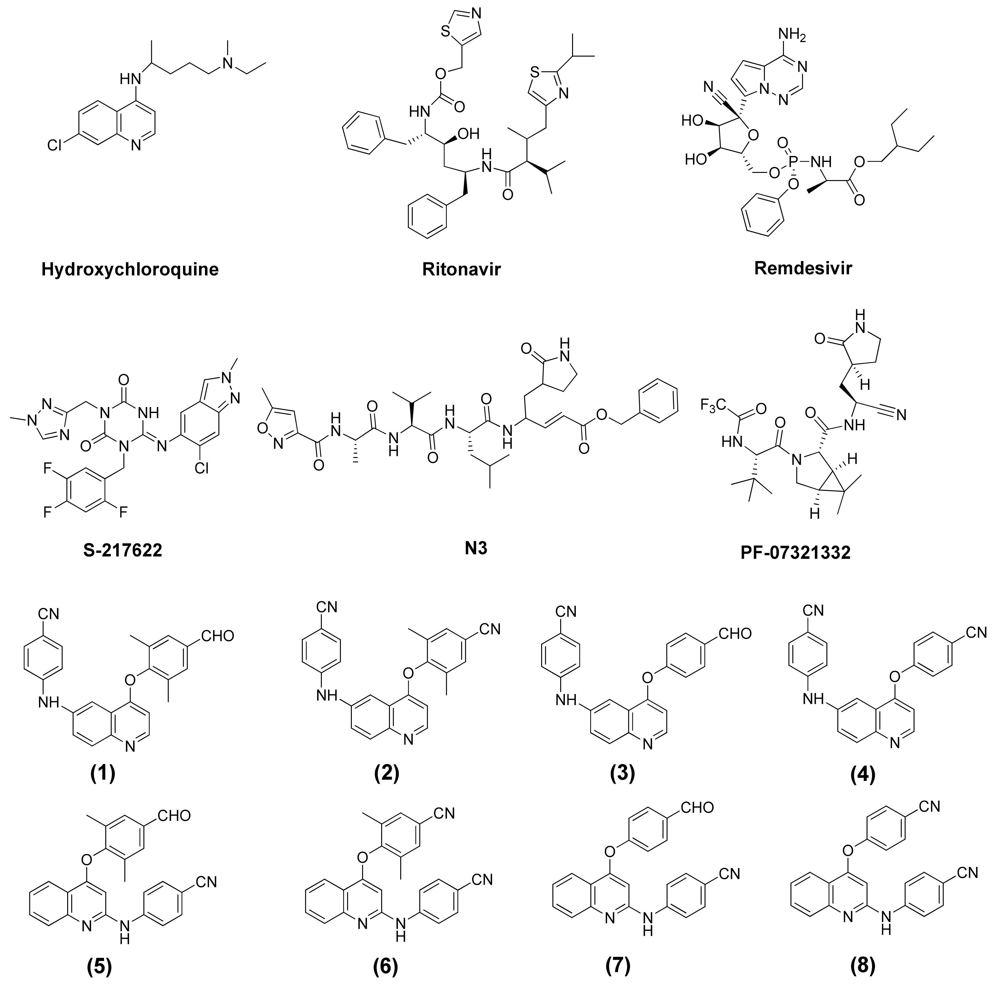
Figure 1. The structure of current medicaments and phenylamino-phenoxy-quinoline derivatives (18).
Molecules 27 01793 g001
Figure 2. (A) The docking conformation of the analyzed ligands in Mpro using molecular docking. (B) Overlaying of the conformations of current medicaments: Hydroxychloroquine (green). Ritonavir (grey), remdesivir (brown), S-217622 (orange), N3 (pink), PF-07321332 (blue), and interesting compounds (14) (red) and (58) (yellow) in the binding pocket of Mpro.
Molecules 27 01793 g002
Figure 3. Overlay of the conformations of the analyzed ligands (green): hydroxychloroquine (A), ritonavir (B), remdesivir (C), S-217622 (D), N3 (E), (1) (F), (6) (G), and (8) (H) with PF-07321332 (blue) in the binding pocket of Mpro.
Molecules 27 01793 g003
Figure 4. Adopted model system of ligands bound to the binding site of Mpro.
Molecules 27 01793 g004
Table 1. Molecular properties of ligands calculated by SwissADME software.
LigandNumber of H-Bond
Acceptors
Number of H-Bond
Donors
LogPNumber of Rotatable BondsMolecular Weight (g/mol)TPSA
2)
Hydroxychloroquine322.359335.8748.39
Ritonavir741.8022720.94202.26
Remdesivir1240.1814602.58213.36
S-217622913.705531.88120.68
N3950.3822680.79197.83
PF-07321332830.4111499.53131.40
(1)412.775393.4475.01
(2)412.775390.5281.73
(3)412.355365.1475.01
(4)412.354362.6581.73
(5)413.175393.4275.01
(6)413.174390.4481.73
(7)412.755365.2975.01
(8)412.754362.3881.73
Table 2. Binding interaction between ligands with Mpro in 5.0 Å.
LigandBinding
Energy
(kcal/mol)
H-BondPi-Pi
Stacking
abc
Hydroxychloroquine−7.06 ± 0.11 LEU141 GLY143 CYS145 HIS164ASN142-HIS41
Ritonavir−8.56 ± 0.25ASN142 GLY143 CYS145 GLN189GLU166 MET165HIS163-
Remdesivir−8.63 ± 0.25GLY143 CYS145 LEU167ASN142 GLU166-HIS41
S-217622−9.62 ± 0.08TYR54 CYS145 GLU166 GLN189PHE140MET165HIS41
N3−9.44 ± 0.16PHE140 GLY143 GLU166HIS41 SER144--
PF-07321332−10.61 ± 0.12PHE140 SER144 CYS145 HIS163 HIS164 GLU166 THR190GLN189--
(1)−10.12 ± 0.01HIS164 GLN192MET49 PRO52 PRO168GLU166HIS41
(2)−9.75 ± 0.08HIS164 GLN192MET49 PRO52GLU166HIS41
(3)−9.67 ± 0.04HIS164 GLN192MET49 PRO52 THR190GLU166HIS41
(4)−9.65 ± 0.03HIS164 GLN192MET49 PRO52-HIS41
(5)−9.72 ± 0.04GLN189 GLN192ASP187GLU166HIS41
(6)−10.02 ± 0.01GLN189 GLN192MET49 ALA191GLU166HIS41
(7)−9.78 ± 0.05HIS164 GLN192MET49 PRO52 PRO168GLU166HIS41
(8)−9.97 ± 0.02CYS145 HIS164 GLN192MET49 PRO52GLU166HIS41
a: Conventional hydrogen bond. b: Carbon hydrogen bond. c: Pi-donor hydrogen bond.
Table 3. Distance between the ligands (current medicaments, 1, 6 and 8) and Mpro within 5.0 Å from the molecular docking.
Amino Acid ResidueHydroxychloroquineRitonavirRemdesivirS-217622N3PF-07321332(1)(6)(8)
HIS414.2083.9544.1584.4573.4122.2954.1783.8973.758
MET49------2.2562.2252.185
PRO52------2.694-2.806
PHE140---3.4452.8152.489---
LEU1412.015--- ----
ASN1423.4211.8992.892- ----
GLY1432.9152.4542.914-2.203----
SER1442.036----2.725---
CYS1452.6142.7013.7253.170-2.245--3.697
HIS163-----1.877---
HIS1642.299----2.1672.104-2.054
MET165-3.0393.1563.053-----
GLU166-2.9642.9372.9432.089/2.2632.012/2.192/2.9542.9622.4322.937
PRO168------2.321--
GLN189-1.984-2.546-2.352-3.141-
THR190-----2.585---
ALA191-------2.910-
GLN192-----2.8381.9541.8722.251
Table 4. Interaction energies of ligands with the individual amino acid residues of Mpro, calculated at the B3LYP/6-31G (d,p) level.
Amino AcidInteraction Energy
(kcal/mol)
HydroxychloroquineRitonavirS-217622N3PF-07321332(1)(6)(8)
HIS41−1.06−0.16−2.32−3.42−0.36−2.06−2.18−2.2
MET490.64−0.08−0.41−0.23−0.87−1.04−1.08−1.47
LEU500.06−0.210.10−0.050.01−0.670.19−0.43
ASN51−0.11−0.140.080.030.06−0.42−0.06−0.26
PRO520.87−0.140.740.01−0.03−0.96−1.01−1.86
ASN53−0.090.070.050.040.12−0.28−0.07−0.26
TYR54−0.09−0.27−3.450.010.071.091.010.9
PHE1401.16−0.21−2.32−4.32−3.610.22−0.16−0.47
LEU141−2.71−0.321.042.3−0.77−0.62−0.880.02
ASN142−1.36−1.01−1.01−0.36−1.210.460.770.21
GLY143−3.14−2.45−1.25−5.02−0.590.20.250.05
SER1440.43−0.18−0.24−3.37−3.96−0.020.41−0.07
CYS145−2.47−2.4−2.89−2.5−3.44−0.67−0.98−2.87
HIS163−0.78−1.69−1.14−2.08−3.05−0.57−0.49−0.66
HIS164−3.2−1.07−1.330.15−2.75−2.98−0.55−3.16
MET1652.290.94−1.890.030.130.481.531.5
GLU166−9.51−18.44−22.49−23.14−24.99−14.47−21.33−21.75
LEU167−0.94−0.82−0.22−0.64−0.320.090.170.11
PRO168−0.420.250.10−0.08−0.05−1.09−0.03−0.83
HIS172−0.19−0.19−0.67−2.28−0.210.26−0.18−0.76
ASP1872.57−0.260.220.49−1.03−0.44−0.430.02
ARG188−3.20.330.98−0.450.49−0.31−1.17−0.57
GLN189−0.44−3.03−3.17−0.08−3.54−1.36−2.15−1.07
THR190−0.221.481.010.070.21−0.85−0.310.42
ALA191−0.18−0.060.020.090.05−0.36−1.19−0.78
GLN192−0.88−0.410.210.070.13−5.31−3.38−3.66
Total−22.97−30.47−40.25−44.73−49.51−31.68−33.30−39.90
Publisher’s Note: MDPI stays neutral with regard to jurisdictional claims in published maps and institutional affiliations.

