Article

Long-Term Evolution of Malnutrition and Loss of Muscle Strength after COVID-19: A Major and Neglected Component of Long COVID-19

1
Transversal Nutrition Unit, University of Lorraine, Nancy University Hospital, 54500 Vandoeuvre-les-Nancy, France
2
Internal Medicine and Clinical Immunology Department, University of Lorraine, Nancy University Hospital, 54500 Vandoeuvre-les-Nancy, France
3
Medical Evaluation Department, Department of Clinical Research Support PARC, University of Lorraine, Nancy University Hospital, 54500 Vandoeuvre-les-Nancy, France
*
Author to whom correspondence should be addressed.
Nutrients 2021, 13(11), 3964; https://doi.org/10.3390/nu13113964
Submission received: 1 September 2021 / Revised: 29 October 2021 / Accepted: 3 November 2021 / Published: 6 November 2021
(This article belongs to the Special Issue Medical Nutrition Therapy in Critically Ill and COVID-19 Patients)

Abstract

:
Post-acute consequences of COVID-19, also termed long COVID, include signs and symptoms persisting for more than 12 weeks with prolonged multisystem involvement; most often, however, malnutrition is ignored. Method: The objective was to analyze persistent symptoms, nutritional status, the evolution of muscle strength and performance status (PS) at 6 months post-discharge in a cohort of COVID-19 survivors. Results: Of 549 consecutive patients hospitalized for COVID-19 between 1 March and 29 April 2020, 23.7% died and 288 patients were at home at D30 post-discharge. At this date, 136 of them (47.2%) presented persistent malnutrition, a significant decrease in muscle strength or a PS ≥ 2. These patients received dietary counseling, nutritional supplementation, adapted physical activity guidance or physiotherapy assistance, or were admitted to post-care facilities. At 6 months post-discharge, 91.0% of the 136 patients (n = 119) were evaluated and 36.0% had persistent malnutrition, 14.3% complained of a significant decrease in muscle strength and 14.9% had a performance status > 2. Obesity was more frequent in patients with impairment than in those without (52.8% vs. 31.0%; p = 0.0071), with these patients being admitted more frequently to ICUs (50.9% vs. 31.3%; p = 0.010). Among those with persistent symptoms, 10% had psychiatric co-morbidities (mood disorders, anxiety, or post-traumatic stress syndrome), 7.6% had prolonged pneumological symptoms and 4.2% had neurological symptoms. Conclusions: Obese subjects as well as patients who have stayed in intensive care have a higher risk of functional loss or undernutrition 6 months after a severe COVID infection. Malnutrition and loss of muscle strength should be considered in the clinical assessment of these patients.

1. Introduction

Post-acute consequences of COVID-19, also termed long COVID, include signs and symptoms that develop during or after an infection consistent with COVID-19, and persist for more than 12 weeks with prolonged multisystem involvement and significant disability. The most commonly described symptoms are sensory (loss of taste and anosmia), neurological (loss of concentration and “brain fog”) and cardiorespiratory problems (fatigue, dyspnea, reduced exercise capacity). In most instances, malnutrition is left ignored as these patients are often overweight at the time of diagnosis [1].
Recent studies have reported a high prevalence of malnutrition among hospitalized patients with COVID-19 depending on the screening and diagnostic tool used [2], and levels are estimated at about 50% (31.7–66.5%) [2]. This prevalence furthermore appears particularly high in patients requiring a stay in intensive care [1,3]. Several factors could explain malnutrition during this acute phase, including marked systemic inflammation driving hypermetabolism and muscle catabolism, and prolonged periods of bedrest driving disuse atrophy. Up to 40% of patients with COVID-19 experience gastrointestinal symptoms ranging from nausea, vomiting, anorexia, diarrhea and abdominal distention, especially in ICU COVID patients [4], which can further deter eating and impact the tolerance of nutritional support [5]. Olfactory and gustatory dysfunction [6] may also contribute to weight loss [7,8].
Muscle loss and/or loss of muscle function appear to be the major nutritional challenges during this acute period. Both myalgia and muscle loss have been strongly correlated with disease severity among COVID-19 patients [9]. Protein turnover is also increased in critical illness in the early stages of COVID-19, in response to massive proteolytic stimuli [10]. Suggested mechanisms include direct muscle invasion by ECoV particles and immune-mediated muscle injury, presenting as myositis, although satisfactory proof of the direct invasion of SARS-CoV-2 into muscle cells is still lacking [11]. Lastly, muscle deconditioning due to immobility and corticosteroid treatment has been associated with diffuse atrophy at muscle biopsy [12]. A retrospective case study from China revealed that 10.7% of patients showed skeletal muscle injury during this acute phase as well as various neurological manifestations (36.4%) [13].
As a result, studies have mainly focused on the importance of nutritional intervention during the acute phase of COVID-19 in order to prevent clinical deterioration (review in [14]). However, the consequences of unintentional weight loss and resulting sarcopenia can also have major long-term functional impacts [15]. A large number of patients are being discharged from hospital following COVID-19 without a systematic assessment of their recovery and need for rehabilitation or further investigation to detect complications, in particular functional complaints related to loss of muscle strength, persistence of malnutrition and fatigue [16]. Medical teams have become increasingly aware of the importance of multidisciplinary care, taking into account fatigue and functional disability, but nevertheless overlooking the nutritional aspect and the importance of the decrease in muscle strength [1]. Nowadays, muscle weakness and fatigue appear as a frequent complaint among these patients [4,5].
In clinical practice, impairment of muscle strength is difficult to assess since, unlike weight, there is no objective assessment of muscle strength prior to disease. Such objective evaluation can be quite straightforward, i.e., using the grip test or chair lifting test. However, these tests are not easily implemented in the context of severe infection or in the patient’s home, and do not establish whether the alteration is linked to the disease or whether it existed beforehand. In view of the latter, we found it useful to use a self-evaluation muscle strength scale, asking the patient how he/she rated his/her muscle strength compared to before the disease. In a preliminary study, we found a concordance between the outcome of this subjective functional evaluation (Self Evaluation of Strength (SES)) and that of the grip test [17].
During the first wave of the pandemic, we rapidly established a post-COVID follow-up service collecting data to identify unmet health needs and to identify those requiring additional rehabilitation and/or investigation for complications. All of these patients received dietary counseling and adapted physical activity guidance, some of whom with severe disability and being re-admitted to post-care facilities. The others received physiotherapy assistance and nutritional supplementation.
The aim of this study was to evaluate, at 6 months post-discharge, the evolution of health status in the group of patients who had a persistent impairment at day 30: namely, patients with persistent weight loss (>5%) or impaired muscle strength (SES < 7/10), or with a Performance Status (PS) > 2. The objective was to analyze the frequency and nature of their symptoms persisting since the initial infection, their nutritional status as well as the evolution of their muscle strength and performance status.

2. Methods

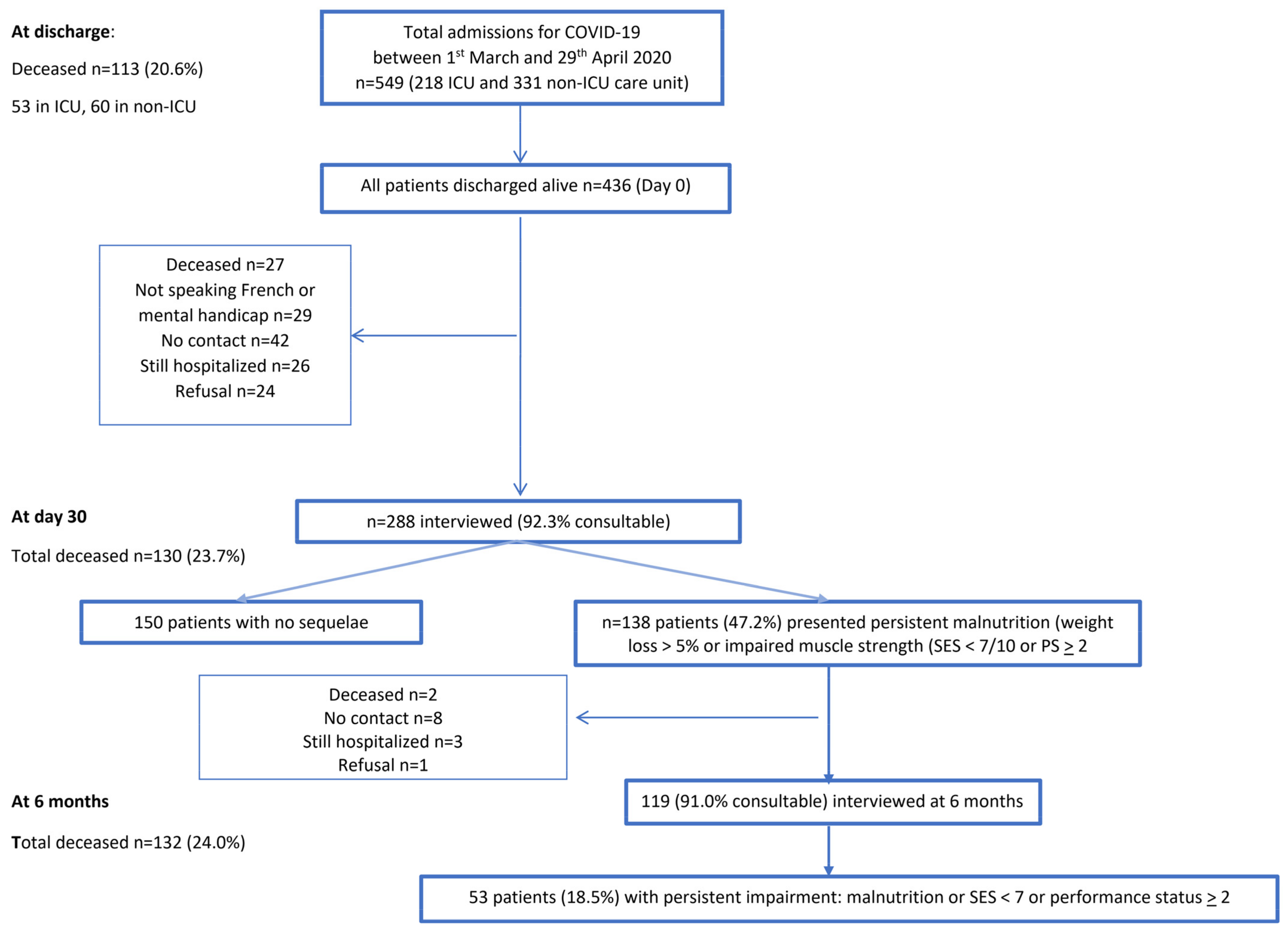
2.1. Study Design and Participants

This study, conducted as part of a prospective cohort study, included all adult inpatients (≥18 years old) who were diagnosed with COVID-19 and admitted to an ICU or non-ICU unit for COVID-19 patients at the Nancy Brabois University Hospital between 1 March 2020 and 29 April 2020 and subsequently discharged alive from hospital. The diagnosis of COVID-19 was based on a positive SARS-CoV-2 RT-PCR test on a nasopharyngeal sample and/or on a typical chest CT scan [18]. The study was approved by the Research Commission of the University Hospital of Nancy and the requirement for informed consent was waived by the ethics commission. The ClinicalTrials.gov (accessed on 16 August 2021) identifier is NCT04451694.
To manage the flow of incoming patients, a certain number were transferred to other hospitals after a few days. At discharge from the intensive or acute care unit, some patients were admitted to post-acute care facilities for ongoing skilled nursing care and rehabilitation.
All hospital-discharged patients were offered a teleconsultation 30 days after discharge (D30) to assess their nutritional status and muscle function, the degree of disability linked to the degree of malnutrition and subjective functional loss, as well as limitation of daily activity estimated by the WHO performance status score. Consequently, patients with persistent weight loss (>5%) at D30 or with impaired muscle strength (Self Evaluation of Strength < 7/10) or a performance status (PS) ≥ 2 (group of impairment patients) were invited to be evaluated 6 months after discharge (by teleconsultation or in person).

2.2. Demographics, Comorbidities and Hospitalization Data Collection

Patient characteristics and hospitalization data were collected by manual review of electronic medical records. Epidemiological, demographic, laboratory and outcome data were extracted from electronic medical records during hospitalization. Sociodemographic data included age, sex, living alone or with others, occupational activity (active vs. unemployed and retired), smoking status (active or not) and daily alcohol consumption. Health characteristics included comorbidities (hypertension, diabetes mellitus, cerebrovascular disease, cardiovascular disease, chronic lung disease) as well as COVID-19 symptoms (anosmia and dysgeusia, diarrhea, dyspnea, asthenia, food aversion).
Hospitalization characteristics included: ICU admission (yes/no), time between symptom onset and hospitalization (days) and length of stay (days).

2.3. Recorded Symptoms

Asthenia at discharge, at day 30 and at 6 months post-discharge was evaluated using a fatigue visual analogue scale (VAS) (0–10) [19].
Dyspnea at discharge, at day 30 and at 6 months post-discharge was evaluated by the French adaptation of the American Thoracic Society Scale, according to 5 levels (from 1 to 5) [20].
Health status at day 30 and at 6 months was assessed using the WHO/Zubrod Performance Status Scale which rates patients from 0 to 4 [21].
Neurological symptoms were also explored (neuropathy, headache, impaired memory and concentration or cognitive impairment).
Depression, anxiety and PTSD diagnoses were based on DSM-V criteria [22,23].

2.4. Nutritional Assessment

Nutritional status prior to hospitalization, on admission, at discharge, at day 30 and at 6 months post-discharge was assessed using anthropometric measurements (BMI: body mass index = body weight/height2) and weight loss (%) compared to weight prior to illness. In hospital, patients were weighed and their height was measured. On day 30 and at 6 months post-discharge, patients were instructed to use their own weighing scales.

2.5. SEFI and Self-Assessment of Muscle Function (SES) at Discharge, at Day 30 and at 6 Months Post-Discharge

Food intake was assessed using the 10-point verbal (AVeS) or visual (AViS) analogue scales (self-evaluation of food intake (SEFI)) [24], graded from 0 to 10. As suggested by Bouette et al. [24], an SEFI < 7 was considered as the cut-off value.
Self-assessment of strength (SES) was assessed at discharge, at day 30 and at 6 months post-discharge using the 10-point verbal (AVeS) or visual (AViS) analogue scales via teleconsultation for evaluating arm and leg strength in comparison to patient strength prior to hospitalization. In practice, patients were asked to evaluate their arm and leg strength in comparison with their strength prior to COVID-19. As suggested by Krznaric et al. [14], patients were asked about their degree of difficulty in lifting or carrying a weight, walking across the room, rising from a chair or bed, and to evaluate these difficulties using a 10-point verbal or visual analogue scale (10 = same strength as before illness and 0 = total loss of strength).
Assessment of physical activity was carried out by completing the International Physical Activity Questionnaire-Short Form (IPAQ-SF) [25] in order to estimate activity prior to COVID-19, with activity classified as low, moderate and high physical activity [25].

2.6. Malnutrition Diagnosis

Malnutrition diagnosis was made according to the GLIM criteria and French recommendations (at least 1 phenotypic criterion and 1 etiological criterion) [26,27]. Severe malnutrition was defined following the French recommendations as weight loss > 10% of weight before COVID-19 infection or a BMI < 17 (<18.5 for patients > 70 years old). Moderate malnutrition was defined as weight loss > 5% of weight before COVID-19 or BMI < 18.5 (<21 for patients > 70 years old) [27,28].

2.7. Statistical Analysis

Continuous variables are expressed as mean ± SD and categorical variables as absolute values and percentages. A paired Student’s t-test, chi-square (χ2), ANOVA, Fisher’s exact test, and Wilcoxon tests were used to compare the values of variables between groups as appropriate. Pearson’s chi-square test or Fisher’s exact test was used to assess the association between each of the discrete variables and the impairment status. p < 0.05 was considered statistically significant. Data were recorded on Excel files. Statistical analysis was performed using Statistical Analysis Software 9.4, SAS Institute Inc, Cary, NC, USA.

3. Results

Population description (Table 1 and Figure 1).
Of the 549 consecutive patients hospitalized for COVID-19 between 1 March and 29 April 2020, 23.7% died and 288 patients were at home at D30 post-discharge (Figure 1).
The mean age of the 288 interviewed patients was 59.8 ± 16.6 years, 54.2% of whom were male. The majority had a low physical activity assessed by IPAQ-SF prior to contracting COVID-19 (56.3%). Seventy-six percent of these patients were overweight or obese. According to the GLIM criteria, 20.7% were already undernourished on admission. The mean delay between the first symptom and admission was 9.7 ± 8.6 days. The most frequent comorbidities were cardiovascular diseases (hypertension, dyslipidemia, diabetes mellitus and coronary artery disease). At admission, anosmia was present in 42.7% of patients, dyspnea in 22.6%, cough in 25.0% and diarrhea in 9.3% of patients.
As reported previously [17], 13.2% of the patients hospitalized for a severe form of COVID-19 were still severely malnourished 30 days after hospital discharge (weight loss > 10% and/or BMI < 17). The highest predictive factors of persistent malnutrition were ICU stay and male sex. Moreover, 26.3% of these patients complained of impaired muscle strength and had a subjective functional loss evaluated at <7/10 using a 10-point verbal (AVeS) or visual (AViS) analogue scale. Lastly, 8.3% (n = 24) concomitantly exhibited a performance status (PS) ≥ 2, severe malnutrition and subjective functional loss.
At D30, 136 patients (47.2%) presented an impairment with persistent malnutrition or impaired muscle strength (SES < 7/10) or severe disability with a PS ≥ 2. Of the latter, 119 (91.0%) accepted phone or teleconsultations or in-person interviews at 6 months. Two patients died during this period, three were still hospitalized, eight were unreachable and there was one refusal (Figure 1).
The evolution of the characteristics of the 119 patients with impairment at D30 is shown in Table 2.
As shown in Table 2, patients with impairment at D30 regained an average of 3.6 kg body weight between D30 and 6 months, with mean weight and BMI returning to near baseline values (admission); on average, patients remained 1.4 kg lighter than on admission. Forty-three patients (36.0% of the 119 patients with impairment at D30) displayed persistent malnutrition at 6 months, with 18 patients exhibiting severe malnutrition (15.1%). On average, seventeen patients (14.3% of the 119 patients with impairment at D30) complained of a significant decrease in muscle strength (SES < 7/10 with a mean ± SD = 5.0 ± 2.6 for arm and 4.9 ± 2.6 for legs). Seventeen patients also had a performance status ≥ 2 (14.9% of the 119 patients with impairment at D30). SES increased between D30 and 6 months post-discharge, nearing the index value prior to COVID infection.

3.1. Long COVID Symptoms at 6 Months Post-Discharge

The most frequent symptoms were asthenia (16.0% had fatigue score > 5/10 at 6 months) and psychiatric disorders; 12 patients (10.0%) experienced mood disorders, anxiety, or post-traumatic stress syndrome. Nine patients (7.6%) presented prolonged pneumological symptoms (dyspnea), and five patients (4.2%) had neurological symptoms (neuropathy, headache, impaired memory and concentration or cognitive impairment).

3.2. Characteristics of Patients with Impairment at 6 Months Post-Discharge, Comparison with Recovered Patients

Overall, at 6 months, 53 patients presented a persistent impairment, as assessed by malnutrition and/or SES < 7 or performance status ≥ 2. The mean characteristics of this population with persistent impairment are described in Table 3 and were compared with the recovered patients (at discharge or at D30).
Comorbidity prevalence did not significantly differ between these two groups, except for obesity which was more frequent in the group with impairment at 6 months post-discharge (52.8% vs. 31.0%; p = 0.007), with these patients being admitted more frequently to ICUs (50.9% vs. 31.3%; p = 0.010). These patients had more difficulties eating after discharge (SEFI: 6.1 ± 3.0 vs. 7.4 ± 2.9 p = 0.0032) and exhibited greater weight loss, with an increased prevalence of malnutrition. SES scores were lower at discharge in this group (4.3 ± 2.4 vs. 5.3 ± 2.6 for arms; p = 0.013, and 4.0 ± 2.0 vs. 5.2 ± 2.7 for legs; p = 0.0045) with no other significant difference being observed.

4. Discussion

This is the first report on a prospective observational cohort study of COVID-19 specifically exploring nutritional status, subjective functional loss and disability at 6 months after discharge. We showed that, at D30, 138/288 of these patients (48%) presented persistent malnutrition (33%), subjective functional loss (26.3%) and/or performance status ≥ 2 (24.3%). At 6 months, 15% of the initial cohort remained malnourished despite nutritional counseling during hospitalization and ensuing dietary guidance, oral nutritional supplements, or relocation to rehabilitation centers, 6% complained of a significant decrease in muscle strength and 6% had a performance status > 2, or 18.5% of this cohort.
Contrary to our results, a study conducted in a cohort of severe COVID-19 patients assessing nutritional status 3 months after discharge from hospital [29] reported that only 8.4% of patients were malnourished at discharge and none at 3 months. Malnutrition was correlated with severity of the disease, indirectly inferable from an assessment of length of hospital stay and need for admission to ICU. However, in this latter study, malnutrition diagnosis was based on the 2015 ESPEN consensus [30], including different criteria for malnutrition than those of the international consensus (GLIM criteria [26]), in particular the postulation that patients with a BMI above 22 are not malnourished. There are several arguments against this latter assertion. The first is that patients suffering from obesity have, because of metabolic modifications (insulin resistance, fatty acid metabolism, hyperglycemia, etc.), higher protein catabolism than subjects of normal weight in the event of traumatism [31], which was also reported in acute COVID patients with obesity [32]. Secondly, accelerated muscle loss is a major factor of morbidity and mortality in obese COVID-19 patients [33]. On the contrary, in our study, obese subjects had a higher risk of functional loss or undernutrition 6 months after a severe COVID infection, as well as patients with a stay in intensive care.
In our cohort, 20.7% of patients were already undernourished at admission. Malnutrition tended to have begun during the initial phases of the disease occurring at home since, upon admission, patients declared significant involuntary weight loss when compared to their habitual weight [29]. However, it is not excluded that malnutrition may precede the infection. A recent study showed that patients with a recent history of malnutrition could be at higher risk of severe COVID-19 [34]. The majority of our patients had comorbidities that could lead to malnutrition. However, we did not observe any influence of these comorbidities on the evolution of their nutritional status. Weight loss and undernutrition were seemingly associated very early with a major decrease in food intake, which remained strongly impaired in half of the patients at hospital discharge, despite nutritional support. Caccialanza et al. [35] also showed that this reduced self-reported food intake prior to hospitalization and/or expected by physicians in the days after admission was associated with negative clinical outcomes in non-critically ill, hospitalized COVID-19 patients. The other component is the increased energy expenditure secondary to a major inflammatory syndrome that leads to hypercatabolism which, associated with reduced food intake and immobilization, significantly contributes to muscle atrophy and sarcopenia.
Acute sarcopenia may mostly affect patient prognosis and incur post-COVID-19 functional and physical deterioration. The degree of muscle mass and functional loss can be influenced by a multiplicity of factors, including the patient’s general pre-infection medical and functional condition, especially in older adults [10]. However, this functional condition prior to infection is typically not analyzed and/or is unknown. The subjective assessment of muscle strength by an analogue or numeric scale could therefore be useful to follow the evolution of muscle strength in these patients. Some patients still appeared very weak 6 months after the infection, despite advice to increase physical activity with protein support, or referral to physiotherapists or adapted physical activity therapists. Most of our patients had low physical activity assessed by IPAQ-SF prior to contracting COVID-19 (56.3%), which may represent a risk factor for severe COVID-infection [36,37].
Fifteen percent of patients presented neuro-psychiatric symptoms requiring treatment. These disorders are well-described in the literature and could be linked to a direct action of the virus on the nervous system [38,39].
Among the strengths and weaknesses of this analysis, the subjective evaluation of muscle strength is subject to debate. The strong point of such evaluation is to assess muscle function in the long term and to judge the evolution of muscle strength, without knowledge of the level of strength prior to the disease. We evaluated this tool in a small cohort of hospitalized patients [17] and while we were unable to identify the actual threshold of functional impairment, our assessment was nevertheless based on the threshold determined according to the questionnaire of Krznaric et al. [14] and on other subjective visual or numeric scales [40]. This threshold must, however, be validated in a large cohort study, notably in comparison with the evolution of muscle strength objectively measured by dynamometry.
Weighing patients at home is also a weak point due to the lack of weight scale control. Nevertheless, this measurement error was, a priori, the same for the patient’s usual weight prior to infection.
Lastly, we hypothesized that patients who were not impaired at discharge or who improved their nutritional status and muscle strength between discharge and D30 had little risk of having a worse outcome thereafter and were thus not contacted after 6 months. However, three additional deaths were observed during this latter period, albeit all linked to an underlying disease.
In conclusion, undernutrition and loss of muscle strength are symptoms of long COVID and should be considered in the clinical assessment of these patients. Although there are no current specific treatments for use in patients who have been hospitalized for COVID-19, treatments should focus on nutritional support and rehabilitation exercises whenever possible to prevent long-term disability as a result of acute illness due to COVID-19, as well as on the management of sarcopenia. The data described herein may assist in the identification of patients outside of expected recovery trajectories who could benefit from additional rehabilitation and/or further investigation to detect post-COVID nutritional complications. Of these outlying patients, obese subjects appear particularly at risk, as well as patients who have stayed in intensive care.

Author Contributions

M.G. Conceptualization—Investigation—Software; M.M., A.M., N.M. and N.S. Investigation; R.J. Supervision; P.-L.N.-T. Conceptualization—Formal Analysis—Methodology; D.Q. Conceptualization—Project administration—Formal analysis—Methodology—Supervision—Writing—original draft—Writing—review & editing. All authors have read and agreed to the published version of the manuscript.

Funding

This research received no external funding.

Institutional Review Board Statement

The study was approved by the Research Commission of the University Hospital of Nancy and the requirement for informed consent was waived by the ethics commission.

Data Availability Statement

The data that support the findings of this study are available from the corresponding author upon reasonable request.

Acknowledgments

This study was conducted with the help of the French Society for Clinical Nutrition and Metabolism. Trial participants were thanked for their contribution to this study and for consenting that the data will be published anonymously.

Conflicts of Interest

All authors declare no conflict of interest in relation with this study.

References

  1. Venturelli, S.; Benatti, S.V.; Casati, M.; Binda, F.; Zuglian, G.; Imeri, G.; Conti, C.; Biffi, A.M.; Spada, M.S.; Bondi; et al. Surviving COVID-19 in Bergamo province: A post-acute outpatient re-evaluation. Epidemiol. Infect. 2021, 149, e32. [Google Scholar] [CrossRef] [PubMed]
  2. Abate, S.M.; Chekole, Y.A.; Estifanos, M.B.; Abate, K.H.; Kabthymer, R.H. Prevalence and outcomes of malnutrition among hospitalized COVID-19 patients: A systematic review and meta-analysis. Clin. Nutr. ESPEN 2021, 43, 174–183. [Google Scholar] [CrossRef] [PubMed]
  3. Anker, M.S.; Landmesser, U.; von Haehling, S.; Butler, J.; Coats, A.J.S.; Anker, S.D. Weight loss, malnutrition, and cachexia in COVID-19: Facts and numbers. J. Cachexia Sarcopenia Muscle 2021, 12, 9–13. [Google Scholar] [CrossRef] [PubMed]
  4. Ye, L.; Yang, Z.; Liu, J.; Liao, L.; Wang, F. Digestive system manifestations and clinical significance of coronavirus disease 2019: A systematic literature review. J. Gastroenterol. Hepatol. 2021, 36, 1414–1422. [Google Scholar] [CrossRef]
  5. Liu, R.; Paz, M.; Siraj, L.; Boyd, T.; Salamone, S.; Lite, T.V.; Leung, K.M.; Chirinos, J.D.; Shang, H.H.; Townsend, M.J.; et al. Feeding intolerance in critically ill patients with COVID-19. Clin. Nutr. 2021, 29, S261–S5614. [Google Scholar]
  6. Tong, J.Y.; Wong, A.; Zhu, D.; Fastenberg, J.H.; Tham, T. The Prevalence of Olfactory and Gustatory Dysfunction in COVID-19 Patients: A Systematic Review and Meta-analysis. Otolaryngol. Head Neck Surg. 2020, 163, 3–11. [Google Scholar] [CrossRef]
  7. Roos, D.S.; Oranje, O.J.M.; Freriksen, A.F.D.; Berendse, H.W.; Boesveldt, S. Flavor perception and the risk of malnutrition in patients with Parkinson’s disease. J. Neural Transm. 2018, 125, 925–930. [Google Scholar] [CrossRef] [Green Version]
  8. Li, T.; Zhang, Y.; Gong, C.; Wang, J.; Liu, B.; Shi, L.; Duan, J. Prevalence of malnutrition and analysis of related factors in elderly patients with COVID-19 in Wuhan, China. Eur. J. Clin. Nutr. 2020, 74, 871–875. [Google Scholar] [CrossRef] [Green Version]
  9. Liu, H.; Chen, S.; Liu, M.; Nie, H.; Lu, H. Comorbid Chronic Diseases are Strongly Correlated with Disease Severity among COVID-19 Patients: A Systematic Review and Meta-Analysis. Aging Dis. 2020, 11, 668–678. [Google Scholar] [CrossRef]
  10. Piotrowicz, K.; Gasowski, J.; Michel, J.P.; Veronese, N. Post-COVID-19 acute sarcopenia: Physiopathology and management. Aging Clin. Exp. Res. 2021, 33, 2887–2898. [Google Scholar] [CrossRef]
  11. Wong, D.W.L.; Klinkhammer, B.M.; Djudjaj, S.; Villwock, S.; Timm, M.C.; Buhl, E.M.; Wucherpfennig, S.; Cacchi, C.; Braunschweig, T.; Knuchel-Clarke, R.; et al. Multisystemic Cellular Tropism of SARS-CoV-2 in Autopsies of COVID-19 Patients. Cells 2021, 10, 1900. [Google Scholar] [CrossRef]
  12. Msigwa, S.S.; Wang, Y.; Li, Y.; Cheng, X. The neurological insights of the emerging coronaviruses. J. Clin. Neurosci. 2020, 78, 1–7. [Google Scholar] [CrossRef]
  13. Mao, L.; Jin, H.; Wang, M.; Hu, Y.; Chen, S.; He, Q.; Chang, J.; Hong, C.; Zhou, Y.; Wang, D.; et al. Neurologic Manifestations of Hospitalized Patients With Coronavirus Disease 2019 in Wuhan, China. JAMA Neurol. 2020, 77, 683–690. [Google Scholar] [CrossRef] [Green Version]
  14. Cereda, E.; Clave, P.; Collins, P.F.; Holdoway, A.; Wischmeyer, P.E. Recovery Focused Nutritional Therapy across the Continuum of Care: Learning from COVID-19. Nutrients 2021, 13, 3293. [Google Scholar] [CrossRef]
  15. Seessle, J.; Waterboer, T.; Hippchen, T.; Simon, J.; Kirchner, M.; Lim, A.; Muller, B.; Merle, U. Persistent symptoms in adult patients one year after COVID-19: A prospective cohort study. Clin. Infect. Dis. 2021, 5, ciab611. [Google Scholar]
  16. Adeloye, D.; Elneima, O.; Daines, L.; Poinasamy, K.; Quint, J.K.; Walker, S.; Brightling, C.E.; Siddiqui, S.; Hurst, J.R.; Chalmers, J.D.; et al. The long-term sequelae of COVID-19: An international consensus on research priorities for patients with pre-existing and new-onset airways disease. Lancet Respir. Med. 2021, 17, S2213-2600. [Google Scholar]
  17. Quilliot, D.; Gerard, M.; Bonsack, O.; Malgras, A.; Vaillant, M.F.; Di Patrizio, P.; Jaussaud, R.; Ziegler, O.; Nguyen-Thi, P.L. Impact of severe SARS-CoV-2 infection on nutritional status and subjective functional loss in a prospective cohort of COVID-19 survivors. BMJ Open 2021, 11, e048948. [Google Scholar] [CrossRef]
  18. Cheng, M.P.; Papenburg, J.; Desjardins, M.; Kanjilal, S.; Quach, C.; Libman, M.; Dittrich, S.; Yansouni, C.P. Diagnostic Testing for Severe Acute Respiratory Syndrome-Related Coronavirus 2: A Narrative Review. Ann. Intern. Med. 2020, 172, 726–734. [Google Scholar] [CrossRef] [Green Version]
  19. Hewlett, S.; Hehir, M.; Kirwan, J.R. Measuring fatigue in rheumatoid arthritis: A systematic review of scales in use. Arthritis Rheum. 2007, 57, 429–439. [Google Scholar] [CrossRef] [Green Version]
  20. Society, A.T. Recommended respiratory disease questionnaires for use with adults and children in epidemiologlcal research. Am. Rev. Respir. Dis. 1978, 118, 7–35. [Google Scholar]
  21. Oken, M.M.; Creech, R.H.; Tormey, D.C.; Horton, J.; Davis, T.E.; McFadden, E.T.; Carbone, P.P. Toxicity and response criteria of the Eastern Cooperative Oncology Group. Am. J. Clin. Oncol. 1982, 5, 649–655. [Google Scholar] [CrossRef]
  22. American Psychiatric Association: DSM-V Development. Available online: https://www.psychiatry.org/psychiatrists/practice/dsm/updates-to-dsm-5//updates-to-dsm-5-criteria-text (accessed on 20 July 2021).
  23. American Psychiatric Association. Diagnostic and Statistical Manual of Mental Disorders, 5th ed.; Arlington, V.A.P.A., Ed.; American Psychiatric Association: Washington, DC, USA, 2013; pp. 271–272. [Google Scholar]
  24. Thibault, R.; Goujon, N.; Le Gallic, E.; Clairand, R.; Sebille, V.; Vibert, J.; Schneider, S.M.; Darmaun, D. Use of 10-point analogue scales to estimate dietary intake: A prospective study in patients nutritionally at-risk. Clin. Nutr. 2009, 28, 134–140. [Google Scholar] [CrossRef]
  25. Craig, C.L.; Marshall, A.L.; Sjostrom, M.; Bauman, A.E.; Booth, M.L.; Ainsworth, B.E.; Pratt, M.; Ekelund, U.; Yngve, A.; Sallis, J.F.; et al. International physical activity questionnaire: 12-country reliability and validity. Med. Sci. Sports Exerc. 2003, 35, 1381–1395. [Google Scholar] [CrossRef] [Green Version]
  26. Cederholm, T.; Jensen, G.L.; Correia, M.; Gonzalez, M.C.; Fukushima, R.; Higashiguchi, T.; Baptista, G.; Barazzoni, R.; Blaauw, R.; Coats, A.; et al. GLIM criteria for the diagnosis of malnutrition—A consensus report from the global clinical nutrition community. Clin. Nutr. 2019, 38, 1–9. [Google Scholar] [CrossRef] [Green Version]
  27. HAS. Diagnostic de la Dénutrition de l’Enfant et de l’Adulte. Saint-Denis La Plaine. 2019. Available online: https://www.has-sante.fr/upload/docs/application/pdf/2019-11/reco277_recommandations_rbp_denutrition_cd_2019_11_13_v0.pdf (accessed on 20 July 2021).
  28. Thibault, R.; Quilliot, D.; Seguin, P.; Tamion, F.; Schneider, S.; Déchelotte, P. Nutritional care at hospital during the Covid-19 viral epidemic: Expert opinion from the French-speaking Society for Clinical Nutrition and Metabolism (SFNCM). Nut. Clin. Métab. 2020, 34, 97–104. [Google Scholar] [CrossRef]
  29. Fiorindi, C.; Campani, F.; Rasero, L.; Campani, C.; Livi, L.; Giovannoni, L.; Amato, C.; Giudici, F.; Bartoloni, A.; Fattirolli, F.; et al. Prevalence of nutritional risk and malnutrition during and after hospitalization for COVID-19 infection: Preliminary results of a single-centre experience. Clin. Nutr. ESPEN 2021, 45, 351–355. [Google Scholar] [CrossRef]
  30. Cederholm, T.; Bosaeus, I.; Barazzoni, R.; Bauer, J.; Van Gossum, A.; Klek, S.; Muscaritoli, M.; Nyulasi, I.; Ockenga, J.; Schneider, S.M.; et al. Diagnostic criteria for malnutrition—An ESPEN Consensus Statement. Clin. Nutr. 2015, 34, 335–340. [Google Scholar] [CrossRef]
  31. Jeevanandam, M.; Young, D.H.; Schiller, W.R. Obesity and the metabolic response to severe multiple trauma in man. J. Clin. Invest. 1991, 87, 262–269. [Google Scholar] [CrossRef] [Green Version]
  32. Gualtieri, P.; Falcone, C.; Romano, L.; Macheda, S.; Correale, P.; Arciello, P.; Polimeni, N.; Lorenzo, A. Body Composition Findings by Computed Tomography in SARS-CoV-2 Patients: Increased Risk of Muscle Wasting in Obesity. Int. J. Mol. Sci. 2020, 21, 4670. [Google Scholar] [CrossRef]
  33. Zhang, X.; Xie, X.; Dou, Q.; Liu, C.; Zhang, W.; Yang, Y.; Deng, R.; Cheng, A.S.K. Association of sarcopenic obesity with the risk of all-cause mortality among adults over a broad range of different settings: A updated meta-analysis. BMC Geriatr. 2019, 19, 183. [Google Scholar] [CrossRef]
  34. Kurtz, A.; Grant, K.; Marano, R.; Arrieta, A.; Grant, K., Jr.; Feaster, W.; Steele, C.; Ehwerhemuepha, L. Long-term effects of malnutrition on severity of COVID-19. Sci. Rep. 2021, 11, 14974. [Google Scholar] [CrossRef] [PubMed]
  35. Caccialanza, R.; Formisano, E.; Klersy, C.; Ferretti, V.; Ferrari, A.; Demontis, S.; Mascheroni, A.; Masi, S.; Crotti, S.; Lobascio, F.; et al. Nutritional parameters associated with prognosis in non-critically ill hospitalized COVID-19 patients: The NUTRI-COVID19 study. Clin. Nutr. 2021, 25, S261–S5614. [Google Scholar]
  36. Wu, C.; Chen, X.; Cai, Y.; Xia, J.; Zhou, X.; Xu, S.; Huang, H.; Zhang, L.; Zhou, X.; Du, C.; et al. Risk Factors Associated With Acute Respiratory Distress Syndrome and Death in Patients With Coronavirus Disease 2019 Pneumonia in Wuhan, China. JAMA Intern. Med. 2020, 180, 934–943. [Google Scholar] [CrossRef] [PubMed] [Green Version]
  37. Williamson, E.J.; Walker, A.J.; Bhaskaran, K.; Bacon, S.; Bates, C.; Morton, C.E.; Curtis, H.J.; Mehrkar, A.; Evans, D.; Inglesby, P.; et al. Factors associated with COVID-19-related death using OpenSAFELY. Nature 2020, 584, 430–436. [Google Scholar] [CrossRef]
  38. Ousseiran, Z.H.; Fares, Y.; Chamoun, W.T. Neurological manifestations of COVID-19: A systematic review and detailed comprehension. Int. J. Neurosci. 2021, 27, 1–16. [Google Scholar] [CrossRef]
  39. Majolo, F.; Silva, G.L.D.; Vieira, L.; Anli, C.; Timmers, L.; Laufer, S.; Goettert, M.I. Neuropsychiatric Disorders and COVID-19: What We Know So Far. Pharmaceuticals 2021, 14, 933. [Google Scholar] [CrossRef]
  40. Bouette, G.; Esvan, M.; Apel, K.; Thibault, R. A visual analogue scale for food intake as a screening test for malnutrition in the primary care setting: Prospective non-interventional study. Clin. Nutr. 2020, 40, 174–180. [Google Scholar] [CrossRef]
Figure 1. Flowchart of patients included in the study.
Nutrients 13 03964 g001
Table 1. Characteristics of the whole population (n = 288).
Variablesn and %
Age (years)59.8 ± 16.6
Sex (F/M)132/156 (45.8%/54.2%)
Living alone54 (18.8%)
Couple132 (45.8%)
Family96 (33.3%)
Retirement home6 (2.1%)
BMI Class
18.5–24.969 (24.3%)
25–29.9112 (39.4%)
>30103 (36.3%)
IPAQ (prior to COVID-19)
Low162 (56.3%)
Moderate82 (28.5%)
High44 (15.3%)
ICU102 (35.4%)
Active smoking12 (4.2%)
Daily alcohol14 (4.9%)
Comorbidities
HBP113 (39.2%)
Coronary heart disease41 (14.2%)
Dyslipidemia64 (22.2%)
Diabetes mellitus48 (16.7%)
Renal failure23 (8.0%)
Stroke9 (3.1%)
Asthma14 (4.9%)
Apnea19 (6.6%)
Chronic obstructive bronchitis13 (4.5%)
Respiratory failure10 (3.5%)
Active cancer30 (10.4%)
Neurological disease13 (4.5%)
BMI, body mass index; IPAQ, International Physical Activity Questionnaire; ICU, intensive care unit; HBP, high blood pressure.
Table 2. Characteristics and evolution of nutritional status at 6 months in the sub-group of patients with impairment at day 30 after severe COVID-19 (n and %).
Admission (n = 119)At Discharge
(n = 119)
D30 Home
(n = 119)
At 6 Months
(n = 119)
p
Weight (kg)82.6 ± 19.0676.5 ± 16.177.6 ± 15.981.2 ± 17.9<10−3
BMI28.7 ± 5.926.7 ± 5.027.4 ± 5.028.3 ± 5.5<10−3
Weight variation (%)−3.7 ± 4.9−5.1 ± 5.9−7.4 ± 5.0−3.6 ± 5.9<10−3
Malnutrition <10−3
No68/119 (57.1%)23/119 (19.3%)22/119 (18.5%)76/119 (63.9%)
Moderate21/119 (17.6%)42/119 (35.3%)43/119 (36.1%)25/119 (21.0%)
Severe12/119 (10.1%)54/119 (45.4%)54/119 (45.4%)18/119 (15.1%)
SEFI 7.2 ± 3.09.6 ± 1.29.8 ± 0.9<10−3
SES hands 4.2 ± 2.46.9 ± 2.19.1 ± 1.7<10−3
SES legs 4.0 ± 2.36.8 ± 2.19.1 ± 1.6<10−3
Subjective functional loss (SES < 7)% 79.8%55.5%14.3%<10−3
Asthenia (VAE)> 5/10 (%) 3.2 ± 3.0
35/119 (29.4%)
1.7 ± 2.5
19/119 (16.0%)
<10−3
0.020
Performance Status <10−3
0 22 (18.5%)30 (25.2%)70/119 (58.8%)
1 44 (37.0%)44 (37.0%)27/119 (22.7%)
2 29 (24.4%)29 (24.4%)9/119 (7.6%)
3–4 24 (20.2%)16 (13.4%)8/119 (6.7%)
D30, day 30; BMI, body mass index; SEFI, self-evaluation of food intake; SES, self-evaluation of strength.
Table 3. Comparison of the characteristics of recovered patients and of the group of patients with impairment at 6 months.
Recovered PatientsPatients with Impairments at 6 Months
VariablesN = 217 (80.4%)N = 53 (19.6%)p
Symptoms at discharge
Dyspnea ≥ 221 (9.6%)3 (5.7%)NS
Anosmia at discharge95 (43.8%)18 (34%)NS
Diarrhea10 (4.6%)2 (3.8%)NS
cough16 (7.4%)4 (7.5%)NS
Obesity (BMI ≥ 30) (%) at admission31.0%52.8%0.007
ICU admission31.3%50.9%0.010
SEFI at discharge7.4 ± 2.96.1 ± 3.00.003
Dietary aversion at discharge19 (8.8%)8 (15.1%)NS
Weight loss (%)
Before admission−2.8% ± 4.1%−5.5 ± 7.00.001
Total at discharge−5.2% ± 4.7%−10.5 ± 6%<0.001
Malnutrition at discharge (%)102 (49.3%)43 (82.6%)0.001
Moderate62 (30.0%)15 (28.8%)
Severe40 (19.3%)28 (53.8%)
SES at discharge
Arms5.3 ± 2.64.3 ± 2.40.013
Legs5.2 ± 2.74.0 ± 2.00.004
SES < 7138 (71.9%)46 (92%)0.003
Fisher’s exact test for qualitative variables, Student’s t test for quantitative variables. SEFI, self-evaluation of food intake; SES, self-evaluation of strength.
Publisher’s Note: MDPI stays neutral with regard to jurisdictional claims in published maps and institutional affiliations.

