Article

Neurological Prognostic Factors in Hospitalized Patients with COVID-19

1
Department of Pharmacology, Jagiellonian University Medical College, 16 Grzegorzecka St., 31-531 Krakow, Poland
2
John Paul II Hospital, 80 Pradnicka St., 31-202 Krakow, Poland
3
Department of Neurology, University Hospital in Krakow, 2 Jakubowskiego St., 30-688 Krakow, Poland
4
Department of Neurology, Jagiellonian University Medical College, 2 Jakubowskiego St., 30-688 Krakow, Poland
5
Department of Infectious Diseases, University Hospital in Krakow, 2 Jakubowskiego St., 30-688 Krakow, Poland
6
Department of Infectious and Tropical Diseases, Jagiellonian University Medical College, 2 Jakubowskiego St., 30-688 Krakow, Poland
7
Department of Otorhinolaryngology, University Hospital in Krakow, 2 Jakubowskiego St., 30-688 Krakow, Poland
8
Department of Otorhinolaryngology, Jagiellonian University Medical College, 2 Jakubowskiego St., 30-688 Krakow, Poland
9
Department of Internal Medicine and Gerontology, Jagiellonian University Medical College, 2 Jakubowskiego St., 30-688 Krakow, Poland
10
Department of Internal Medicine and Gerontology, University Hospital in Krakow, 2 Jakubowskiego St., 30-688 Krakow, Poland
11
Department of Metabolic Diseases and Diabetology, Jagiellonian University Medical College, 2 Jakubowskiego St., 30-688 Krakow, Poland
12
Department of Metabolic Diseases and Diabetology, University Hospital in Krakow, 2 Jakubowskiego St., 30-688 Krakow, Poland
13
Department of Neurology, University of Rochester Medical Center, Rochester, 601 Elmwood Ave, Rochester, NY 14642, USA
*
Author to whom correspondence should be addressed.
Brain Sci. 2022, 12(2), 193; https://doi.org/10.3390/brainsci12020193
Submission received: 28 November 2021 / Revised: 27 January 2022 / Accepted: 28 January 2022 / Published: 30 January 2022
(This article belongs to the Collection COVID-19 and Brain)

Abstract

:
We aimed to search whether neurological symptoms or signs (NSS) and the MEWS (Modified Early Warning Score) score were associated with in-hospital mortality or oxygen requirement during the first 14 days of hospitalization in COVID-19 patients recruited at the University Hospital in Krakow, Poland. The detailed clinical questionnaires on twenty NSS were either filled out by patients prospectively or retrospectively assessed by neurologists based on daily medical records. NSS were considered high or low-risk if they were associated with increased or decreased mortality in the univariable analysis. This cohort study included 349 patients with COVID-19 (median age 64, interquartile range (51–77), women 54.72%). The presence of high-risk NSS (decreased level of consciousness, delirium, seizures, and symptoms of stroke or transient ischemic attack) or its combination with the absence of low-risk NSS (headache, dizziness, decreased mood, and fatigue) increased the risk of in-hospital mortality in SARS-CoV-2 infection 3.13 and 7.67-fold, respectively. The presence of low-risk NSS decreased the risk of in-hospital mortality in COVID-19 patients more than 6-fold. Death in patients with SARS-CoV-2 infection, apart from NSS, was predicted by older age, neoplasm, and higher MEWS scores on admission. High-risk NSS or their combination with the absence of low-risk NSS increased the risk of oxygen requirement during hospitalization in COVID-19 patients 4.48 and 1.86-fold, respectively. Independent predictors of oxygen therapy during hospitalization in patients with SARS-CoV-2 infection were also older age, male sex, neoplasm, and higher MEWS score on admission.

1. Introduction

The Severe Acute Respiratory Syndrome coronavirus 2 (SARS-CoV-2) infection caused a significant burden for public health systems, with over 5.5 million deaths worldwide as of 22 January 2022 [1]. Mortality among patients admitted to hospital due to this disease was found to be nearly three times higher when compared to hospitalized cases with influenza [2]. Additionally, more than half of young patients with Coronavirus Disease 2019 (COVID-19) required oxygen therapy during hospitalization [3]. The COVID-19 pandemic also resulted in the increased incidence of mental health disorders, such as anxiety, depression, and insomnia [4]. The broad variety of neurological symptoms in the course of SARS-CoV-2 infection ranged from mild manifestations, including myalgia, headache [5], fatigue, dizziness, and anosmia, to more severe presentations, such as seizures, stroke, and encephalopathy [6]. Involvement of both the central and peripheral nervous systems was common within the first 14 days of the SARS-CoV-2 infection [7]. Moreover, a substantial number of patients experienced persistent neurological symptoms within 90 days after discharge from the COVID-19 ward [8].
Therefore, the need for simple and reliable prognostic factors that could be helpful in predicting the course of SARS-CoV-2 infection during hospitalization still exist [9]. Previous studies pointed to many clinical [10] and paraclinical [11] findings as possible risk factors for deterioration of patient or death. For example, in an Italian study of 87 patients with COVID-19, a high Monocyte Distribution Width (MDW) value emerged as the prognostic factor of fatal outcome with sensitivity 0.75 and specificity 0.70 [12] that reflected hyperinflammation mediated by monocyte/macrophage subsets [13,14,15,16]. In another study, the presence of neurological symptoms that were associated with a poorer prognosis was predicted by low lymphocyte count and increased lactate dehydrogenase and interleukin-6 [17]. However, a recent German study showed that self-reported patients’ symptoms after COVID-19 corresponded well with the objective findings in the neuropsychological testing, including fatigue, sleep disturbances, anxiety, and depression, especially among hospitalized patients [18]. Moreover, neurological manifestations became increasingly noted in the course of COVID-19, even in critically ill patients [19], and some of them, such as cerebrovascular disease, were associated with worse prognoses [20]. Our previous study revealed that different neurological symptoms increased in-hospital mortality, whereas others exhibited a protective role in this issue [21]. However, the role of the combination of different neurological symptoms in mediating the risk of death or requirement for oxygen therapy was not sufficiently investigated.
Many prognostic scales corresponded with mortality risk among patients with COVID-19, such as Pneumonia Severity Index [22] or CURB-65 [23], among others. In Poland, where more than 4 million cases with SARS-CoV-2 infection were noted as of 22 January 2022 [1], the easy to obtain Modified Early Warning Score (MEWS) was extensively used in clinical practice in accordance with national COVID-19 guidelines presented at the beginning of the pandemic [24]. However, there is limited data on the prognostic significance of the MEWS score in patients with COVID-19, and available studies produced divergent results [25,26,27].
Therefore, the aim of the current study was to search whether the combination of easy to identify neurological symptoms, prior central nervous system (CNS) diseases, including stroke and MEWS score, was associated with in-hospital mortality or the requirement of oxygen therapy among hospitalized patients with COVID-19.

2. Materials and Methods

2.1. Patients

This cohort study included 349 patients with COVID-19 hospitalized between March 2020 and February 2021 at five departments of the University Hospital in Krakow, Poland: Neurology (42.69%, n = 149), Metabolic Diseases and Diabetology (31.23%, n = 109), Infectious Diseases (16.62%, n = 58), Internal Medicine (5.73%, n = 20), and Otorhinolaryngology (3.72%, n = 13). The SARS-CoV-2 infection was diagnosed based on the positive result of real-time reverse transcription-polymerase chain reaction (97.99%, n = 342) or antigen test (2.01%, n = 7) from a nasopharyngeal swab. The following inclusion criteria were used in the study, being the same as causes of hospitalization: low blood saturation (≤92%), dyspnea, chronic disease that needed hospital treatment, or no possibility for social isolation. We excluded patients below the age of 18 and those who required mechanical ventilation and intubation during hospital admission (cardiopulmonary arrest, lost airway or jeopardized airway, respiratory distress with respiratory rate >30/min, or hypoxemia SpO2 < 93% on room air and PaO2:FiO2 < 300 mmHg that progressively became worse besides 2-hour high-flow oxygen therapy [28]).
We gathered the data on basic demographics, concomitant chronic diseases with special emphasis on cardiovascular and neurological disorders, COVID-19 symptomatology, and blood parameters previously associated with poorer COVID-19 prognosis, which were troponin I [29] and D-dimer levels [30]. Central nervous system diseases included the following: the history of stroke, epilepsy, prior traumatic brain injury, tumor, parkinsonian syndrome, and dementia. All data were gathered retrospectively in a specially designed database that was fulfilled by neurologists based on medical records in the hospital system.

2.2. Neurological Symptoms and Sings

The presence of neurological symptoms in the course of SARS-CoV-2 infection was evaluated prospectively (59.03%, n = 206) or retrospectively (40.97%, n = 143) as described previously [21]. In brief, the detailed clinical questionnaires on the presence of 20 neurological symptoms or signs (NSS), including 12 symptoms (headache, dizziness, decreased mood, memory or concentration difficulties, fatigue, visual disturbances, anosmia, ageusia, muscle weakness, myalgia, paresthesia, increased sweating) and 8 signs (decreased level of consciousness, delirium, ataxia, seizures, stroke/TIA, autonomic disturbances defined as diarrhea, arterial hypotension <90/60 mmHg or tachycardia >100/min), were either filled out by patients prospectively during the first 14 days of hospitalization or retrospectively assessed by neurologists based on daily medical records in the hospital database. Neurological symptoms or signs, such as headache, dizziness, decreased mood, or fatigue, if not reported by patients, were considered absent. Neurological symptoms and signs that were associated with increased mortality in the univariable analysis were considered high-risk NSS, whereas those that decreased the risk of death in the same analysis were defined as low-risk NSS. From such differentiation of NSS excluded were arterial hypotension and tachycardia as these parameters were dependent on the hemodynamic state of the patient that in turn was reflected by MEWS score on admission. The main study endpoints were the requirement of oxygen therapy or death during hospitalization.
Each patient followed prospectively gave informed medical consent that was either written or verbal in the presence of two witnesses. The study was approved by the Bioethics Committee of the District Medical Council in Krakow (opinion number 143/KBL/OIL/2020) and conducted in accordance with the Declaration of Helsinki.

2.3. Statistics

We assumed a 25% difference in the prevalence of NSS based on Brieghel et al. [31]. For such a difference or greater, a minimum sample size of 27 patients in each group was required to achieve an α level of 0.05 and statistical power of 0.80. Patients were stratified according to outcome (death, oxygen therapy) and a group of NSS. We presented categorical values as counts and percentages, and continuous values as means ± standard deviations (SD) or medians (interquartile ranges, IQR), as appropriate. We assessed the distribution normality of continuous variables with the Shapiro–Wilk test and compared, by Student’s t-test or by Mann–Whitney U test, as appropriate. Categorical variables were assessed with a chi-square test. To identify predictors of death and oxygen therapy, we used multivariable logistic regression with backward stepwise elimination. All variables that showed association with outcome in the univariable model with p < 0.05 and showed no substantial correlation with other independent variables (r > 0.5) were included in a model. Non-linear variables were log-transformed. Models for death included age, prior CNS disease, diabetes mellitus, chronic kidney disease stage ≥3, neoplasm, MEWS score, troponin I, D-dimer, and (1) high-risk NSS (model A), (2) low-risk NSS (model B), and (3) high-risk/or absence of low-risk NSS (model C). The multivariable models (1–3) for oxygen therapy also included hypertension and excluded chronic kidney disease stage ≥3. To assess the goodness of fit, Akaike information criterion and the Wald test were assessed. For each model, the area under the receiver operating characteristic curve (AUC) with 95% confidence intervals (CI) was used to assess predictive accuracy. For each group of NSS, AUC with 95% CI, accuracy, sensitivity, specificity, positive and negative predictive value together with positive likelihood ratio was assessed. Probability values ≤0.05 were considered statistically significant. The data were evaluated using STATISTICA (version 13.0, Statsoft Inc., Tulsa, OK, USA).

3. Results

The data that confirm the results of the current study are available from the corresponding author upon reasonable request.

3.1. Patient Characteristics
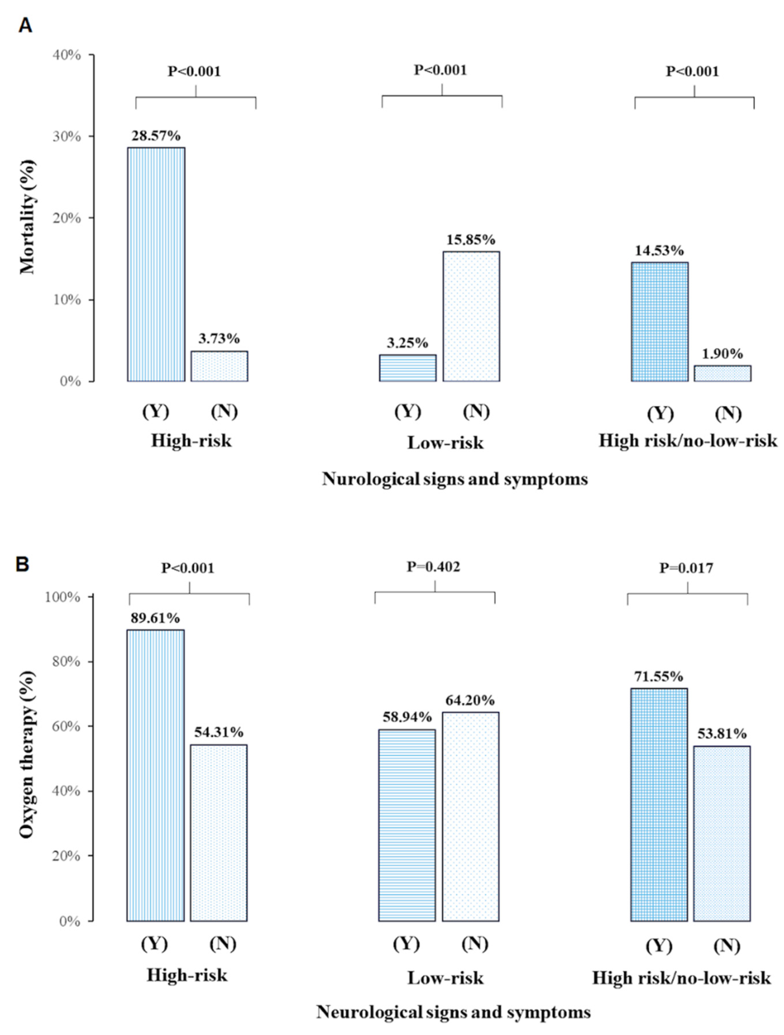
The cohort of 349 patients (median age 64, interquartile range (51–77), women 54.72%) due to COVID-19 is presented in Table 1. Patients were admitted from home (60.74%, n = 212), other hospitals (31.52%, n = 110), nursing homes (6.88%, n = 24), and institutional isolation (0.86%, n = 3). The median time from the first positive nasopharyngeal swab test for SARS-CoV-2 to hospitalization was 1 (1–3) day, and the median time from the onset of COVID-19 symptoms to hospital admission was 5 (3–8) days.

3.2. Mortality

Patients who died during hospitalization (9.46%, n = 33) were older, had a higher prevalence of diabetes mellitus, prior CNS disease, neoplasm, chronic kidney disease stage 3, and higher MEWS score, troponin I, and D-dimer compared to survivors (Table 1). Based on the univariable analysis concerning the risk of death, high-risk NSS comprised the following: a decreased level of consciousness, delirium, seizures, and symptoms of stroke or transient ischemic attack (TIA). Conversely, low-risk NSS included headache, dizziness, decreased mood, and fatigue that were reported by patients (Table S1). High-risk NSS were nearly 4-fold more frequent, and low-risk NSS were more than 2-fold less frequent in this group than in COVID-19 survivors (Table 1, Figure 1A). These results were similar when we separately analyzed patients assessed prospectively and retrospectively; however, there were more deaths in the group collected retrospectively (see Figure S1). There was also accordance with the findings of the study coming from the whole group when patients from different departments were analyzed separately according to the presence of high-risk NSS (see Figure S2).
In the multivariable regression model, death was predicted by age, neoplasm, MEWS score, high-risk NSS, and prior CNS disease (model A), or low-risk NSS (model B), or high-risk/or absence of low-risk NSS (model C) (Table 2). Model C was the best-fitted model based on AIC values. The AUC of high-risk NSS and low-risk NSS for predicting death was 0.756 (95% CI 0.659–0.853) and 0.697 (95% CI 0.572–0.822), respectively (Table S2).

3.3. Oxygen Therapy

Patients requiring oxygen therapy during hospitalization (n = 218, 62.46%) were older and more often males, had a higher prevalence of hypertension, diabetes mellitus, neoplasm, prior CNS disease, and had higher MEWS score, troponin I, and D-dimer compared with the remainder. On admission, they were characterized with a higher frequency of fever, cough, and dyspnea, and lower frequency of sore throat. On day 14 since the onset of hospitalization, 62 (17.12%) patients required oxygen therapy, including 13 (3.72%) mechanically ventilated. We observed a more than five-fold increase in high-risk NSS during hospitalization in the group requiring oxygen therapy (Table 1, Figure 1B).
The independent predictors of oxygen therapy during hospitalization were age, male sex, neoplasm, MEWS score, and high-risk NSS (model A) or high-risk / or absence of low-risk NSS (model C) (Table 3). Model A yielded the lowest AIC values. The AUCs of high-risk NSS for predicting oxygen therapy was 0.630 (95% CI: 0.572–0.689) (Table S2).

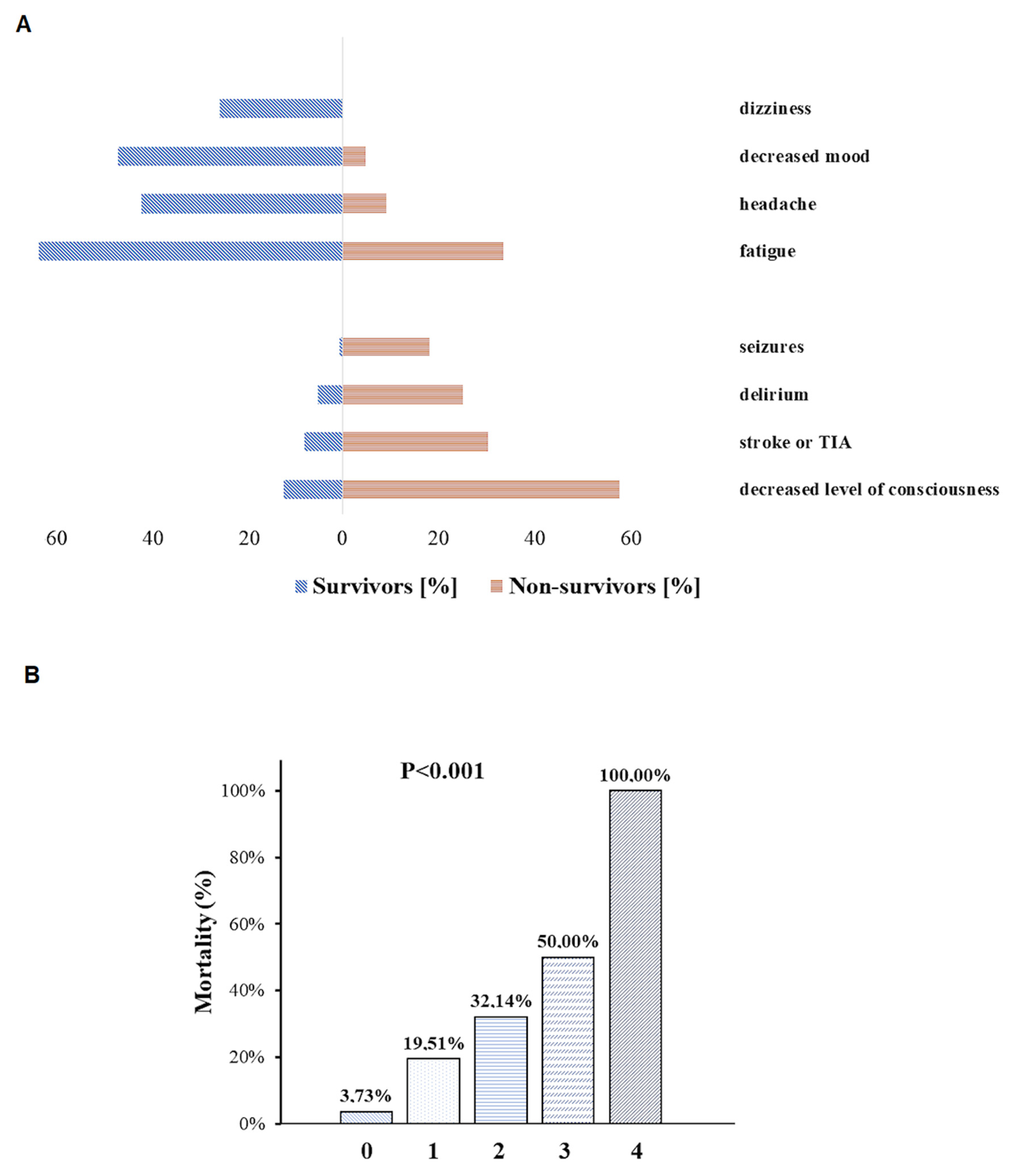
3.4. The Severity of Neurological Symptoms and Signs

3.4.1. High-Risk NSS

Almost one in four (n = 77, 22.06%) patients had one or more high-risk NSS, namely decreased level of consciousness (n = 57, 16.33%), delirium (n = 24, 6.88%), seizures (n = 8, 2.30%), and stroke or TIA symptoms (n = 35, 10.03%) (Table S1, Figure 2A). There were 36 patients with more than one high-risk NSS, including n = 28 (7.98%) with two, n = 6 (1.72%) with three, and n = 2 (0.57%) with four high-risk NSS, respectively.
Patients with high-risk NSS were older (76 (66–84) vs. 61 (49–71) years, p < 0.001), had a higher prevalence of hypertension (73.52 vs. 55.22%, p = 0.002), diabetes mellitus (40.26 vs. 22.01%, p = 0.001), ischemic heart disease (28.57 vs. 13.43%, p = 0.002), prior CNS disease (63.64 vs. 17.72%, p < 0.001), tachycardia (40.26 vs. 26.87%, p = 0.024), and arterial hypotension (45.45 vs. 11.19%, p < 0.001), a higher requirement for oxygen therapy with a non-re-breather mask on admission (46.75 vs. 10.48%, p < 0.001); higher MEWS score (MEWS ≥3, 13.33 vs. 2.61%, p < 0.001), troponin I (13.98 (5.80–33.47) vs. 5.00 (2.72–11.88) mg/dL, p < 0.001), and D-dimer (1.45 (0.72–3.23) vs. 0.63 (0.41–1.23) mg/L, p < 0.001) compared to the remainder (Table S1). The prevalence of sore throat (5.19 vs. 13.93%, p = 0.024) and loss of appetite (7.80 vs. 27.79%, p < 0.001) as a first COVID-19 symptom was lower in the group with high-risk NSS (Table S1).
Patients with any high-risk NSS had higher mortality compared with those who did not manifest symptoms (28.57 vs. 3.73%, p < 0.001) (Figure 1A). The presence of at least one high-risk NSS was associated with more than a 3-fold increase in the risk of death in the multivariable model (Table 2). With the increasing number of high-risk NSS, the prognosis deteriorated, with 50% or more risk of death when three or four symptoms coexisted in one patient (Figure 2B). Any high-risk NSS were associated with an increased need for oxygen therapy (88.31 vs. 54.31%, p < 0.001) and borderline with high-concentration oxygen therapy via a non-rebreather mask (46.75% vs. 10.48%, p = 0.08) compared with patients without those symptoms (Table S1, Figure 1B). Stroke or TIA during COVID-19 analyzed as a single NSS was not associated with the risk of death (OR = 2.59, 95% CI: 0.78–8.65, p = 0.122) or oxygen therapy requirement (OR = 2.09, 95% CI: 0.74–5.93, p = 0.164) in the multivariable analyses (see Tables S3 and S4).

3.4.2. Low-Risk NSS

A total of 246 patients (70.49%) had at least one low-risk NSS: headache (n = 130, 37.24%), dizziness (n = 78, 22.35%), decreased mood (n = 44, 41.26%), and fatigue (n = 200, 57.31%). More than one low-risk NSS was found in 187 (53.58%) patients (Table S1, Figure 2A).
Patients with low-risk NSS were younger (61 (49–73) vs. 70 (57–79) years, p = 0.008), had a lower prevalence of hypertension (55.69 vs. 69.51%, p = 0.027), prior CNS disease (13.41 vs. 30.49%, p < 0.001), lower troponin I (5.46 (2.79–12.94) vs. 10.18 (4.53–21.94) mg/dL, p = 0.001), and D-dimer (0.66 (0.43–1.27) vs. 1.00 (0.43–1.82) mg/L, p = 0.042) compared with the remainder. The prevalence of asthma/chronic obstructive pulmonary disease (7.32 vs.1.22%, p = 0.041), fever (63.01 vs. 48.78%, p = 0.024), cough (67.07 vs. 54.88%, p = 0.047), sore throat (15.04 vs. 6.10%, p = 0.036), loss of appetite (38.62 vs. 10.98%, p < 0.001), dyspnea (54.07 vs. 35.37%, p = 0.034), and abdominal pain (23.58 vs. 8.54%, p = 0.003), as first COVID-19 symptoms, was higher in the low-risk NSS group (Table S1).
Patients with any low-risk NSS had lower mortality (3.25 vs. 15.85%, p < 0.001) compared with those who did not have those symptoms or were unable to report them (Figure 1A). The presence of at least one low-risk NSS was associated with more than a 6-fold decrease in the risk of death in the multivariable model but not oxygen therapy (Table 2 and Table 3).

4. Discussion

To our knowledge, the current study is the first to show that a combination of neurological symptoms, rather than a single symptom, may predict in-hospital mortality in patients with COVID-19. We found that a combination of high-risk NSS and the absence of low-risk NSS, increased the risk of in-hospital death nearly 8-fold in patients with SARS-CoV-2 infection. It was previously noted that isolated neurological symptoms, such as decreased level of consciousness [32], delirium [33], or stroke symptoms [34], were associated with increased mortality in COVID-19 patients. The latest literature review showed that any neurological manifestation of SARS-CoV-2 infection was correlated with a higher mortality rate [35]. A similar conclusion came from a prospective study of Egyptian patients with COVID-19, demonstrating a significant association between neurological events and both a severe course of the disease at onset and higher mortality [7]. On the other hand, in an Italian study of 901 COVID-19 patients, after adjustment for age, sex, and comorbidities, isolated neurological symptoms increased the chance of survival, but nearly one-third of these symptoms comprised dysgeusia, anosmia, or syncope [36]. Of these, anosmia was associated with a nearly 7-fold decrease in the risk of death among more than 11,000 patients with COVID-19 included in a large meta-analysis of 26 studies [37]. A recent Brazilian retrospective cohort study that included 613 patients with COVID-19 showed that, apart from older age and a requirement for mechanical ventilation, patients with encephalopathy had a significantly higher risk of death, but those with anosmia had a lower risk of dying [6]. Many other studies confirmed the protective role of another neurological symptom, namely headache, in terms of the risk of death due to SARS-CoV-2 infection [21,38,39]. Therefore, it appears that different NSS exhibit a diverse correlation with the mortality risk in COVID-19 patients. Moreover, not only does the quality of these symptoms but also their quantity play an important role, with more high-risk NSS, including seizures, delirium, decreased level of consciousness, or stroke or TIA symptoms, leading to an increased risk of death in COVID-19 patients. What is worth emphasizing is that our models for evaluating high and low-risk NSS were also accurate based on AIC/AUC ratios with high negative predictive values.
Our study also revealed that high-risk NSS were independent predictors of oxygen therapy requirement during hospitalization. The literature to date regarding this issue is sparse. A previous study including nearly 900 patients from the NHS Trust hospital in London reported that the main indicators of poor outcomes in COVID-19 were age and oxygenation status in the emergency room [40]. An analysis of the Brazilian Ministry of Health Database encompassing hospitalized cases with SARS-CoV-2 infection showed that blood oxygen saturation levels below 95% increased the risk of death by 1.27-fold [41]. On the other hand, in a Korean study of more than 5000 patients with COVID-19, logistic regression analyses showed that male sex and older age increased the risk of oxygen therapy requirements during hospitalization [42], similar to the findings in our study. A recent analysis from one of the Indian tertiary centers revealed that patients with SARS-CoV-2 infection who died in the course of the disease received high flow nasal cannula oxygen nearly four times more often than survivors [43].
Our study demonstrates the important prognostic role of the MEWS score in patients with COVID-19 since a score of three or more points was associated with a higher risk of death or oxygen demand during hospitalization in each statistical model tested. Patients with respiratory and systemic symptoms of COVID-19 with a MEWS score lower than three were perceived as stable according to previous national guidelines [24]. These findings were similar to our study, in which nearly one-fourth of patients with SARS-CoV-2 also had prior CNS disease. However, previous studies on the MEWS score as a prognostic factor in COVID-19 patients produced contradictory results. A small Chinese study showed that the REMS (Rapid Emergency Medicine Score), which included, in addition, age and oxygenation, correlated better with mortality risk than the MEWS score [44]. A larger Chinese study comparing five different early warning scores in COVID-19 patients showed that the MEWS, when measured on admission, did not predict death risk [25]. On the other hand, a Turkish study revealed that the MEWS score calculated in the emergency room effectively predicted death in the forthcoming 28 days in patients with COVID-19 who required hospitalization [26]. The results of this study were in line with findings from a large cohort of 1000 patients from five Dutch hospitals [45]. A retrospective analysis of data from Wuhan showed that the MEWS was accurate in predicting in-hospital mortality in elderly patients with SARS-CoV-2 infection [46]. A small Polish study also showed that the MEWS predicted a severe course of SARS-CoV-2 infection [47]. The MEWS, which appears to be reliable and easy to assess, was found to be useful in predicting neurological worsening in a cohort of more than 7000 neurocritically ill patients with COVID-19 [48]. However, a recent Indian study showed that not many rating scales, including the MEWS, were found to be useful in identifying patients with COVID-19 who are at risk for clinical worsening [27].
In our study, we were able to show that patients with COVID-19 and prior CNS disease had more than a five-fold increase in the risk of in-hospital mortality. A previous study performed in a group of nearly 600 COVID-19 patients from four hospitals in Ohio found that, although a pre-existent major neurological disease, such as dementia, stroke, or epilepsy, increased the risk of death by a factor of two, it was not an independent predictor of mortality in the multivariate analysis [49]. The same conclusions could be drawn from a small Korean study of COVID-19 patients in an intensive care unit where a history of neurological disease did not affect mortality, in contrast to new-onset neurological complications [50]. On the other hand, the presence of chronic neurological comorbidities, such as a prior stroke with long-term sequelae, cognitive disorders, or neuromuscular and spinal diseases, independently predicted the risk of death in a Spanish cohort of patients with SARS-CoV-2 infection [51]. Most of our cohort with prior CNS disease consisted of patients with pre-existent stroke. A history of stroke was found to independently predicate in-hospital mortality in a large cohort of more than 3000 patients with COVID-19 hospitalized in the Mount Sinai Health System in New York [52]. Similarly, in a Romanian study where nearly 8% of COVID-19 patients had a prior stroke, the presence of a preexisting neurological disorder was highly correlated with SARS-CoV-2 infection severity [53].
Our study has important limitations. First, the number of patients was rather small. The cohort of patients was mixed, and only 59% prospectively filled out clinical questionnaires. Second, information on low-risk NSS was obtained only voluntarily from our patients. Third, selection bias was present in some patient cohorts, particularly in the Departments of Otorhinolaryngology and Internal Medicine, where cohort sizes were small. There were also a small number of patients with mild SARS-CoV-2 infection in the first wave of the pandemic who were hospitalized only due to the lack of the ability to socially isolate. Fourth, all patients in our study were Caucasian; therefore, the results might not be generalizable to other ethnicities, as was shown in a large meta-analysis [54]. Fifth, measuring serum [12] or cerebrospinal fluid (CSF) [55] biomarkers could potentially improve the prognostic ability of our COVID-19 models, but based on the AIC/AUC values, our predictive models appear to be accurate. Sixth, neuroimaging, CSF analyses, and electroencephalography were performed only in the minority of our patients and guided by the clinical history and neurological examination. Previous studies reported that certain neuroimaging features, such as cerebral microbleeds or leukoencephalopathy, were associated with higher mortality and worse outcome on discharge [56]. Therefore, future studies are needed to answer the question of whether additional testing such as neuroimaging could improve our ability to predict the course of COVID-19.

5. Conclusions

The presence of high-risk NSS (decreased level of consciousness, delirium, seizures, and symptoms of stroke or transient ischemic attack) or its combination with the absence of low-risk NSS (headache, dizziness, decreased mood, and fatigue) increases the risk of in-hospital mortality in SARS-CoV-2 infection 3- and nearly 8-fold, respectively. The presence of low-risk NSS decreases the risk of in-hospital mortality in COVID-19 patients more than 6-fold. Death in patients with SARS-CoV-2 infection, apart from NSS, is predicted by older age, neoplasm, and higher MEWS scores on admission. High-risk NSS or the combination of high-risk or absence of low-risk NSS increases the risk of oxygen requirement during hospitalization in COVID-19 patients nearly 5- and 2-fold, respectively. Independent predictors of oxygen therapy during hospitalization in patients with SARS-CoV-2 infection are also older age, male sex, neoplasm, and higher MEWS score on admission.

Supplementary Materials

The following are available online at https://www.mdpi.com/article/10.3390/brainsci12020193/s1, Table S1: Patients’ characteristics according to severity of neurological symptoms and signs, Table S2: Diagnostic accuracy for predicting in-hospital deaths or oxygen therapy, Table S3: Multivariable regression analysis for death with stroke/TIA symptoms, Table S4: Multivariable regression analysis for oxygen therapy with stroke/TIA symptoms, Figure S1: Mortality rates and requirement for oxygen therapy in COVID-19 patients according to the severity of neurological symptoms and signs and method of data collection, Figure S2: Mortality rates and requirement for oxygen therapy in COVID-19 patients according to the severity of neurological symptoms and signs and hospital department.

Author Contributions

L.D., conceptualization, formal analysis, draft writing; J.D., data acquisition, draft writing; Z.C.-B., data acquisition; I.M., data acquisition; K.S., data acquisition; T.K., data acquisition; J.C., data acquisition; P.W., data acquisition; J.S., data acquisition; M.W.-B., supervision, draft review; A.G., supervision, draft review; M.M., supervision, draft review; R.J., language proofreading, draft review; A.S., supervision, draft review; M.W., conceptualization, supervision, draft writing. All authors have read and agreed to the published version of the manuscript.

Funding

The study was supported by the grant from the National Centre for Research and Development CRACoV-HHS project (model of multi-specialist hospital and non-hospital care for patients with SARS-CoV-2 infection) through the initiative “Support for specialist hospitals in fighting the spread of SARS-CoV-2 infection and in treating COVID-19” (contract number-SZPITALE-JEDNOIMIENNE/18/2020). The described research was implemented by the consortium of the University Hospital in Krakow and the Jagiellonian University Medical College.

Institutional Review Board Statement

The study was approved by the Bioethics Committee of the District Medical Council in Krakow (opinion number 143/KBL/OIL/2020) and conducted in accordance with the Declaration of Helsinki.

Informed Consent Statement

Each patient followed prospectively gave informed medical consent that was either written or verbal in the presence of two witnesses.

Data Availability Statement

The data that confirm the results of the current study are available from the corresponding author upon reasonable request.

Acknowledgments

The authors would like to thank Pawel Strek for his agreement to perform the study in the Department of Otorhinolaryngology, University Hospital in Krakow. The authors would also like to thank the staff of the University Hospital in Krakow for contributing to this article: Michalina Adamczyk, Szymon Andrasik, Jakub Antczak, Artur Igor Bien, Pawel Bryniarski, Kinga Czerwiec, Mateusz Czyzycki, Mateusz Dwojak, Mateusz Fiema, Agnieszka Fryzlewicz, Elzbieta Gradek-Kwinta, Tomasz Grodzicki, Agnes Hajek, Jerzy Hohendorff, Barbara Kaminska, Michal Kania, Karol Kasprzycki, Barbara Katra, Alicja Kepinska-Wnuk, Elzbieta Klimiec-Moskal, Marianna Kopka, Malgorzata Kostrzycka, Wojciech Kozminski, Jeremiasz Kubisiowski, Jeremiasz Jagiella, Krzysztof Kurowski, Patrycja Lechowicz, Anna Lopatkiewicz, Monika Marona, Maciej Motyl, Malgorzata Napierala, Klaudia Nowak, Olga Nurkowska, Michal Paykart, Lukasz Pawlinski, Ewelina Pietak, Kamila Pospiech, Anna Prosniak, Agnieszka Pulyk, Roman Pulyk, Gabriela Rusin, Monika Rybicka, Agnieszka Rzeminska, Paulina Surowiec, Damian Ucieklak, Jolanta Walczewska, Kamil Wezyk, Magdalena Wilk, Magdalena Witkowska, Ewa Wlodarczyk, Malgorzata Wlodarczyk, Katarzyna Wojcik, Joanna Zieba-Parkitny.

Conflicts of Interest

All the authors report no disclosures and conflict of interest.

References

  1. Xu, B.; Kraemer, M.U.G.; Xu, B.; Gutierrez, B.; Mekaru, S.; Sewalk, K.; Loskill, A.; Wang, L.; Cohn, E.; Hill, S.; et al. Open access epidemiological data from the COVID-19 outbreak. Lancet Infect. Dis. 2020, 20, 534. [Google Scholar] [CrossRef] [Green Version]
  2. Piroth, L.; Cottenet, J.; Mariet, A.-S.; Bonniaud, P.; Blot, M.; Tubert-Bitter, P.; Quantin, C. Comparison of the characteristics, morbidity, and mortality of COVID-19 and seasonal influenza: A nationwide, population-based retrospective cohort study. Lancet Respir. Med. 2021, 9, 251–259. [Google Scholar] [CrossRef]
  3. Ni, Y.N.; Wang, T.; Liang, B.M.; Liang, Z.A. The independent factors associated with oxygen therapy in COVID-19 patients under 65 years old. PLoS ONE 2021, 16, e0245690. [Google Scholar] [CrossRef]
  4. Wańkowicz, P.; Szylińska, A.; Rotter, I. Insomnia, anxiety, and depression symptoms during the COVID-19 pandemic may depend on the pre-existent health status rather than the profession. Brain Sci. 2021, 11, 1001. [Google Scholar] [CrossRef]
  5. Kacprzak, A.; Malczewski, D.; Domitrz, I. Headache Attributed to SARS-CoV-2 Infection or COVID-19 Related Headache—Not Migraine-like Problem-Original Research. Brain Sci. 2021, 25, 1406. [Google Scholar] [CrossRef]
  6. Sampaio Rocha-Filho, P.A.; Magalhães, J.E.; Fernandes Silva, D.; Carvalho Soares, M.; Marenga Arruda Buarque, L.; Dandara Pereira Gama, M.; Oliveira, F.A.A. Neurological manifestations as prognostic factors in COVID-19: A retrospective cohort study. Acta Neurol. Belg. 2022. online ahead. [CrossRef]
  7. Mekkawy, D.A.; Hamdy, S.; Abdel-Naseer, M.; Shehata, H.S.; Halfawy, A.A.; Shalaby, N.M.; Shehata, G.A.; Ali, A.M.; Elmazny, A.; Ahmed, S.M.; et al. Neurological Manifestations in a Cohort of Egyptian Patients with COVID-19: A Prospective, Multicenter, Observational Study. Brain Sci. 2022, 12, 74. [Google Scholar] [CrossRef]
  8. Wong-Chew, R.M.; Rodríguez Cabrera, E.X.; Rodríguez Valdez, C.A.; Lomelin-Gascon, J.; Morales-Juárez, L.; de la Cerda, M.L.R.; Villa-Romero, A.R.; Arce Fernández, S.; Serratos Fernandez, M.; Bello, H.H.; et al. Symptom cluster analysis of long COVID-19 in patients discharged from the Temporary COVID-19 Hospital in Mexico City. Ther. Adv. Infect. Dis. 2022, 9, 204993612110692. [Google Scholar] [CrossRef]
  9. Dickens, B.S.L.; Lim, J.T.; Low, J.W.; Lee, C.K.; Sun, Y.; Nasir, H.B.M.; Akramullah, F.A.B.M.; Yan, G.; Oon, J.; Yan, B.; et al. Simple “Rule-of-6” Predicts Severe Coronavirus Disease 2019 (COVID-19). Clin. Infect. Dis. 2021, 72, 1861–1862. [Google Scholar] [CrossRef]
  10. Rinderknecht, M.D.; Klopfenstein, Y. Predicting critical state after COVID-19 diagnosis: Model development using a large US electronic health record dataset. NPJ Digit. Med. 2021, 4, 113. [Google Scholar] [CrossRef]
  11. Incerti, D.; Rizzo, S.; Li, X.; Lindsay, L.; Yau, V.; Keebler, D.; Chia, J.; Tsai, L. Prognostic model to identify and quantify risk factors for mortality among hospitalised patients with COVID-19 in the USA. BMJ Open 2021, 11, e047121. [Google Scholar] [CrossRef] [PubMed]
  12. Riva, G.; Castellano, S.; Nasillo, V.; Ottomano, A.M.; Bergonzini, G.; Paolini, A.; Lusenti, B.; Milić, J.; De Biasi, S.; Gibellini, L.; et al. Monocyte Distribution Width (MDW) as novel inflammatory marker with prognostic significance in COVID-19 patients. Sci. Rep. 2021, 11, 12716. [Google Scholar] [CrossRef] [PubMed]
  13. Riva, G.; Nasillo, V.; Luppi, M.; Tagliafico, E.; Trenti, T. Linking COVID-19, monocyte activation and sepsis: MDW, a novel biomarker from cytometry. EBioMedicine 2022, 75, 103754. [Google Scholar] [CrossRef] [PubMed]
  14. Siddiqi, H.K.; Mehra, M.R. COVID-19 illness in native and immunosuppressed states: A clinical–therapeutic staging proposal. J. Heart Lung Transplant. 2020, 39, 405–407. [Google Scholar] [CrossRef] [Green Version]
  15. Riva, G.; Nasillo, V.; Tagliafico, E.; Trenti, T.; Luppi, M. COVID-19: Room for treating T cell exhaustion? Crit. Care 2020, 24, 229. [Google Scholar] [CrossRef]
  16. Gibellini, L.; De Biasi, S.; Paolini, A.; Borella, R.; Boraldi, F.; Mattioli, M.; Lo Tartaro, D.; Fidanza, L.; Caro-Maldonado, A.; Meschiari, M.; et al. Altered bioenergetics and mitochondrial dysfunction of monocytes in patients with COVID-19 pneumonia. EMBO Mol. Med. 2020, 12, e13001. [Google Scholar] [CrossRef]
  17. Zhao, T.-S.; Zeng, H.-L.; Zhang, X.; Chen, X.; Jiang, W.-L.; Du, J.; Liu, H.-Y.; Zhao, J.; Yuan, Y.; Peng, X.-F.; et al. Neurological manifestations in COVID-19 patients and their application in predicting fatal disease: A retrospective cohort study. J. Microbiol. Immunol. Infect. 2021. in press. [CrossRef]
  18. Bungenberg, J.; Humkamp, K.; Hohenfeld, C.; Rust, M.I.; Ermis, U.; Dreher, M.; Hartmann, N.K.; Marx, G.; Binkofski, F.; Finke, C.; et al. Long COVID-19: Objectifying most self-reported neurological symptoms. Ann. Clin. Transl. Neurol. 2022. online ahead. [CrossRef]
  19. Deana, C.; Verriello, L.; Pauletto, G.; Corradi, F.; Forfori, F.; Cammarota, G.; Bignami, E.; Vetrugno, L. Insights into neurological dysfunction of critically ill COVID-19 patients. Trends Anaesth. Crit. Care 2021, 36, 30–38. [Google Scholar] [CrossRef]
  20. Vakili, K.; Fathi, M.; Hajiesmaeili, M.; Salari, M.; Saluja, D.; Tafakhori, A.; Sayehmiri, F.; Rezaei-Tavirani, M. Neurological Symptoms, Comorbidities, and Complications of COVID-19: A Literature Review and Meta-Analysis of Observational Studies. Eur. Neurol. 2021, 84, 307–324. [Google Scholar] [CrossRef]
  21. Wnuk, M.; Sawczyńska, K.; Kęsek, T.; Wrona, P.; Chatys-Bogacka, Ż.; Mazurkiewicz, I.; Drabik, L.; Jagiełła, J.; Szaleniec, J.; Czepiel, J.; et al. Neurological symptoms in hospitalised patients with COVID-19 and their association with in-hospital mortality. Neurol. Neurochir. Pol. 2021, 55, 314–321. [Google Scholar] [CrossRef] [PubMed]
  22. Satici, C.; Asim, M.; Sargin, E.; Gursoy, B. Performance of pneumonia severity index and CURB-65 in predicting 30-day mortality in patients with COVID-19. Int. J. Infect. Dis. 2020, 98, 84–89. [Google Scholar] [CrossRef] [PubMed]
  23. García Clemente, M.M.; Herrero Huertas, J.; Fernández Fernández, A.; De La Escosura Muñoz, C.; Enríquez Rodríguez, A.I.; Pérez Martínez, L.; Gómez Mañas, S.; Iscar Urrutia, M.; López González, F.J.; Madrid Carbajal, C.J.; et al. Assessment of risk scores in Covid-19. Int. J. Clin. Pract. 2021, 75, e13705. [Google Scholar] [CrossRef] [PubMed]
  24. Flisiak, R.; Horban, A.; Jaroszewicz, J.; Kozielewicz, D.; Pawłowska, M.; Parczewski, M.; Piekarska, A.; Simon, K.; Tomasiewicz, K.; Zarębska-Michaluk, D. Management of SARS-CoV-2 infection: Recommendations of the Polish Association of Epidemiologists and Infectiologists as of 31 March 2020. Pol. Arch. Intern. Med. 2020, 130, 352–357. [Google Scholar] [CrossRef] [Green Version]
  25. Hu, H.; Yao, N.; Qiu, Y. Predictive value of five early warning scores for critical novel coronavirus disease. Disaster Med. Public Health Prep. 2020, 1–8. [Google Scholar] [CrossRef]
  26. Aygun, H.; Eraybar, S. The role of emergency department triage early warning score (TREWS) and modified early warning score (MEWS) to predict in-hospital mortality in COVID-19 patients. Ir. J. Med. Sci. 2021, 1–7. [Google Scholar] [CrossRef]
  27. Kaeley, N.; Mahala, P.; Kabi, A.; Choudhary, S.; Hazra, A.; Vempalli, S. Utility of early warning scores to predict mortality in COVID-19 patients: A retrospective observational study. Int. J. Crit. Illn. Inj. Sci. 2021, 11, 163. [Google Scholar] [CrossRef]
  28. Meng, L.; Qiu, H.; Wan, L.; Ai, Y.; Xue, Z.; Guo, Q.; Deshpande, R.; Zhang, L.; Meng, J.; Tong, C.; et al. Intubation and Ventilation amid the COVID-19 Outbreak: Wuhan’s Experience. Anesthesiology 2020, 132, 1317–1332. [Google Scholar] [CrossRef] [Green Version]
  29. Chen, H.; Li, X.; Marmar, T.; Xu, Q.; Tu, J.; Li, T.; Han, J.; Xu, D.; Shen, T. Cardiac Troponin I association with critical illness and death risk in 726 seriously ill COVID-19 patients: A retrospective cohort study. Int. J. Med. Sci. 2021, 18, 1474–1483. [Google Scholar] [CrossRef]
  30. Rostami, M.; Mansouritorghabeh, H. D-dimer level in COVID-19 infection: A systematic review. Expert Rev. Hematol. 2020, 13, 1265–1275. [Google Scholar] [CrossRef]
  31. Brieghel, C.; Ellekvist, P.; Lund, M.L.; Søborg, C.; Walsted, E.S.; Thomsen, J.J.; Biering-Sørensen, T.; Mohr, T.; Knop, F.K.; Ravn, P. Prognostic factors of 90-day mortality in patients hospitalised with covid-19. Dan. Med. J. 2021, 68, 1–9. [Google Scholar]
  32. Xiong, W.; Lu, L.; Zhang, B.; Luo, J.; Li, W.; He, L.; Sander, J.W.; Mu, J.; Zhu, C.; Zhou, D. Association of consciousness impairment and mortality in people with COVID-19. Acta Neurol. Scand. 2021, 144, 251–259. [Google Scholar] [CrossRef] [PubMed]
  33. Morandi, A.; Rebora, P.; Isaia, G.; Grossi, E.; Faraci, B.; Gentile, S.; Bo, M.; Valsecchi, M.G.; Deiana, V.; Ghezzi, N.; et al. Delirium symptoms duration and mortality in SARS-CoV2 elderly: Results of a multicenter retrospective cohort study. Aging Clin. Exp. Res. 2021, 33, 2327–2333. [Google Scholar] [CrossRef] [PubMed]
  34. Syahrul, S.; Maliga, H.A.; Ilmawan, M.; Fahriani, M.; Mamada, S.S.; Fajar, J.K.; Frediansyah, A.; Syahrul, F.N.; Imran, I.; Haris, S.; et al. Hemorrhagic and ischemic stroke in patients with coronavirus disease 2019: Incidence, risk factors, and pathogenesis—A systematic review and meta-analysis. F1000Research 2021, 10, 1–18. [Google Scholar] [CrossRef] [PubMed]
  35. Leven, Y.; Bösel, J. Neurological manifestations of COVID-19—An approach to categories of pathology. Neurol. Res. Pract. 2021, 3, 39. [Google Scholar] [CrossRef]
  36. Travi, G.; Rossotti, R.; Merli, M.; D’Amico, F.; Chiappetta, S.; Giussani, G.; Panariello, A.; Corradin, M.; Vecchi, M.; Raimondi, A.; et al. Neurological manifestations in patients hospitalized with COVID-19: A retrospective analysis from a large cohort in Northern Italy. Eur. J. Neurosci. 2021, 53, 2912–2922. [Google Scholar] [CrossRef] [PubMed]
  37. Goshtasbi, K.; Pang, J.; Lehrich, B.M.; Vasudev, M.; Birkenbeuel, J.L.; Abiri, A.; Kuan, E.C. Association Between Olfactory Dysfunction and Critical Illness and Mortality in COVID-19: A Meta-analysis. Otolaryngol.-Head Neck Surg. 2021, 1–5. [Google Scholar] [CrossRef]
  38. Moradzadeh, R.; Jamalian, S.M.; Nazari, J.; Kamali, A.; Sadeghi, B.; Hosseinkhani, Z.; Sofian, M.; Zamanian, M. Age-standardized mortality rate and predictors of mortality among COVID-19 patients in Iran. J. Educ. Heal Promot. 2021, 10, 169. [Google Scholar] [CrossRef]
  39. Amanat, M.; Rezaei, N.; Roozbeh, M.; Shojaei, M.; Tafakhori, A.; Zoghi, A.; Darazam, I.A.; Salehi, M.; Karimialavijeh, E.; Lima, B.S.; et al. Neurological manifestations as the predictors of severity and mortality in hospitalized individuals with COVID-19: A multicenter prospective clinical study. BMC Neurol. 2021, 21, 1–12. [Google Scholar] [CrossRef]
  40. Heldt, F.S.; Vizcaychipi, M.P.; Peacock, S.; Cinelli, M.; McLachlan, L.; Andreotti, F.; Jovanović, S.; Dürichen, R.; Lipunova, N.; Fletcher, R.A.; et al. Early risk assessment for COVID-19 patients from emergency department data using machine learning. Sci. Rep. 2021, 11, 4200. [Google Scholar] [CrossRef]
  41. De Souza, F.S.H.; Hojo-Souza, N.S.; de Oliveira Batista, B.D.; da Silva, C.M.; Guidoni, D.L. On the analysis of mortality risk factors for hospitalized COVID-19 patients: A data-driven study using the major Brazilian database. PLoS ONE 2021, 16, e0248580. [Google Scholar] [CrossRef] [PubMed]
  42. Jang, S.Y.; Seon, J.Y.; Eun, B.L.; Koh, S.B.; Yoo, J.H.; Lee, W.Y.; Yum, H.K.; Yoon, S.J.; Oh, I.H.; Bae, S.C.; et al. Risk Factors of Outcomes of COVID-19 Patients in Korea: Focus on Early Symptoms. J. Korean Med. Sci. 2021, 36, e132. [Google Scholar] [CrossRef] [PubMed]
  43. Sundaramurthy, R.; Balasubramanian, S.; Ganesan, V.; Aggarwal, P.; Parvataneni, T.; Jyothi Ramachandran Nair, D.P.; Saravanan, R.P. Clinical and Laboratory Factors in Predicting Mortality Among COVID-19 RT-PCR Positive Patients: A Retrospective Observational Study From a Tertiary Care Center. Cureus 2021, 13, e19791. [Google Scholar] [CrossRef] [PubMed]
  44. Hu, H.; Kong, W.; Yao, N.; Qiu, Y.; Yao, R. Prognostic value of three rapid scoring scales and combined predictors for the assessment of patients with coronavirus disease 2019. Nurs. Open 2021, 1–8. [Google Scholar] [CrossRef] [PubMed]
  45. Nijman, G.; Wientjes, M.; Ramjith, J.; Janssen, N.; Hoogerwerf, J.; Abbink, E.; Blaauw, M.; Dofferhoff, T.; van Apeldoorn, M.; Veerman, K.; et al. Risk factors for in-hospital mortality in laboratory-confirmed COVID-19 patients in the Netherlands: A competing risk survival analysis. PLoS ONE 2021, 16, e0249231. [Google Scholar] [CrossRef]
  46. Wang, L.; Lv, Q.; Zhang, X.; Jiang, B.; Liu, E.; Xiao, C.; Yu, X.; Yang, C.; Chen, L. The utility of MEWS for predicting the mortality in the elderly adults with COVID-19: A retrospective cohort study with comparison to other predictive clinical scores. PeerJ 2020, 8. [Google Scholar] [CrossRef]
  47. Stachura, T.; Celejewska-Wójcik, N.; Polok, K.; Górka, K.; Lichołai, S.; Wójcik, K.; Krawczyk, J.; Kozłowska, A.; Przybyszowski, M.; Włoch, T.; et al. A clinical profile and factors associated with severity of the disease among Polish patients hospitalized due to COVID-19—An observational study. Adv. Respir. Med. 2021, 89, 124–134. [Google Scholar] [CrossRef]
  48. Hester, J.; Youn, T.S.; Trifilio, E.; Robinson, C.P.; Babi, M.-A.; Ameli, P.; Roth, W.; Gatica, S.; Pizzi, M.A.; Gennaro, A.; et al. The Modified Early Warning Score: A Useful Marker of Neurological Worsening but Unreliable Predictor of Sepsis in the Neurocritically Ill—A Retrospective Cohort Study. Crit. Care Explor. 2021, 3, e0386. [Google Scholar] [CrossRef]
  49. Salahuddin, H.; Afreen, E.; Sheikh, I.S.; Lateef, S.; Dawod, G.; Daboul, J.; Karim, N.; Gharaibeh, K.; Al-Chalabi, M.; Park, S.; et al. Neurological Predictors of Clinical Outcomes in Hospitalized Patients With COVID-19. Front. Neurol. 2020, 11, 1–8. [Google Scholar] [CrossRef]
  50. Park, J.; Kwon, Y.-S.; Kim, H.-A.; Kwon, D.-H.; Hwang, J.; Jang, S.-H.; Park, H.; Sohn, S.-I.; Choi, H.A.; Hong, J.-H. Clinical Implications of Neurological Comorbidities and Complications in ICU Patients with COVID-19. J. Clin. Med. 2021, 10, 2281. [Google Scholar] [CrossRef]
  51. García-Azorín, D.; Martínez-Pías, E.; Trigo, J.; Hernández-Pérez, I.; Valle-Peñacoba, G.; Talavera, B.; Simón-Campo, P.; de Lera, M.; Chavarría-Miranda, A.; López-Sanz, C.; et al. Neurological Comorbidity Is a Predictor of Death in Covid-19 Disease: A Cohort Study on 576 Patients. Front. Neurol. 2020, 11, 1–8. [Google Scholar] [CrossRef] [PubMed]
  52. Kummer, B.R.; Klang, E.; Stein, L.K.; Dhamoon, M.S.; Jetté, N. History of Stroke Is Independently Associated with In-Hospital Death in Patients with COVID-19. Stroke 2020, 51, 3112–3114. [Google Scholar] [CrossRef] [PubMed]
  53. Gogu, A.E.; Motoc, A.G.; Stroe, A.Z.; Docu Axelerad, A.; Docu Axelerad, D.; Pârv, F.; Munteanu, G.; Dan, F.; Jianu, D.C. Clinical spectrum and neuroimagistic features in hospitalized patients with neurological disorders and concomitant coronavirus-19 infection. Brain Sci. 2021, 11, 1138. [Google Scholar] [CrossRef] [PubMed]
  54. Sze, S.; Pan, D.; Nevill, C.R.; Gray, L.J.; Martin, C.A.; Nazareth, J.; Minhas, J.S.; Divall, P.; Khunti, K.; Abrams, K.R.; et al. Ethnicity and clinical outcomes in COVID-19: A systematic review and meta-analysis. EClinicalMedicine 2020, 29–30, 100630. [Google Scholar] [CrossRef] [PubMed]
  55. Jarius, S.; Pache, F.; Körtvelyessy, P.; Jelčić, I.; Stettner, M.; Franciotta, D.; Keller, E.; Neumann, B.; Ringelstein, M.; Senel, M.; et al. Cerebrospinal fluid findings in COVID-19: A multicenter study of 150 lumbar punctures in 127 patients. J. Neuroinflamm. 2022, 19, 19. [Google Scholar] [CrossRef] [PubMed]
  56. Agarwal, S.; Jain, R.; Dogra, S.; Krieger, P.; Lewis, A.; Nguyen, V.; Melmed, K.; Galetta, S. Cerebral Microbleeds and Leukoencephalopathy in Critically Ill Patients with COVID-19. Stroke 2020, 51, 2649–2655. [Google Scholar] [CrossRef] [PubMed]
Figure 1. Mortality rates (panel A) and requirement for oxygen therapy (panel B) in COVID-19 patients according to the severity of neurological symptoms and signs (NSS). High-risk NSS include decreased level of consciousness, delirium, seizures, and symptoms of stroke or transient ischemic attack. Low-risk NSS comprise headache, dizziness, decreased mood, and fatigue.
Brainsci 12 00193 g001
Figure 2. Occurrence of neurological symptoms and signs in COVID-19 survivors and non-survivors (panel A). Mortality rates according to the number of high-risk neurological symptoms and signs (panel B).
Brainsci 12 00193 g002
Table 1. Patients’ demographic and clinical characteristics and association with death and oxygen therapy.
DeathOxygen Therapy
All Patients
n = 349
No
n = 316
Yes
n = 33
p-ValueNo
n= 131
Yes
n = 218
p-Value
Demographics
Age (years)64 (51–77)62 (49–75)77 (73–84)<0.00158 (45–69)68.5 (55–79)<0.001
Age >75 years, n (%)101 (28.94)80 (25.31)21 (63.63)<0.00122 (16.79)80 (36.70)<0.001
Female sex, n (%)191 (54.72)173 (54.75)18 (54.55)0.98285 (64.89)105 (48.17)0.002
Comorbidities and treatment
Hypertension, n (%)209 (59.89)187 (59.18)22 (66.67)0.40365 (49.62)144 (66.06)0.002
Obesity, n (%)65 (18.62)58 (18.35)7 (21.21)0.68820 (15.27)46 (21.10)0.178
Smoking, n (%)54 (15.47)4 (14.92)7 (21.21)0.34217 (12.98)37 (17.13)0.301
Diabetes mellitus, n (%)90 (25.79)76 (24.05)14 (42.42)0.02226 (19.85)64 (29.36)0.049
Ischemic heart disease, n (%)59 (16.91)51 (16.14)8 (24.24)0.23716 (12.21)43 (19.72)0.077
Prior CNS disease, n (%)77 (22.06)56 (17.72)21 (63.64)<0.00117 (12.98)60 (27.52)0.002
Stroke40 (11.46)27 (8.54)13 (39.39)<0.00110 (7.63)30 (13.82)0.079
Dementia19 (5.44)16 (5.06)3 (9.10)0.3320 (0.00)19 (8.76)<0.001
Parkinsonian syndrome7 (2.01)4 (1.23)3 (9.09)0.0292 (1.53)5 (2.30)0.714
Epilepsy13 (3.72)12 (3.80)1 (3.03)1.0004 (3.05)9 (4.15)0.773
CNS tumor8 (2.29)4 (1.27)4 (12.12)0.0041 (0.76)7 (3.23)0.267
Traumatic brain injury5 (1.43)3 (0.95)2 (6.06)0.0721 (0.76)4 (1.84)0.654
Asthma / COPD, n (%)20 (5.73)20 (6.33)0 (0.0)0.2374 (3.05)17 (7.80)0.102
Neoplasm, n (%)40 (11.46)31 (9.81)9 (27.27)0.0276 (4.58)33 (15.14)0.002
Chronic kidney disease stage 3, n (%)17 (4.87)12 (3.80)5 (15.15)0.0393 (2.29)14 (6.42)0.121
Immunosupressive treatment, n (%)18 (5.16)16 (5.06)2 (6.06)0.6835 (3.82)13 (5.96)0.460
First COVID-19 symptoms
Fever, n (%)207 (59.31)188 (59.49)19 (57.68)0.83167 (51.15)141 (64.68)0.012
Cough, n (%)219 (62.75)199 (62.97)20 (60.61)0.78872 (54.96)147 (67.43)0.019
Sore throat, n (%)44 (12.61)42 (13.29)2 (6.06)%0.40524 (18.32)20 (9.17)0.013
Loss of appetite, n (%)105 (30.09)100 (31.65)5 (15.15)0.07139 (29.77)67 (30.73)0.849
Dyspnea, n (%)178 (51.00)156 (49.37)22 (66.67)0.05834 (25.95)145 (66.50)<0.001
Abdominal pain, n (%)67 (19.20)62 (19.62)5 (15.15)0.53522 (16.79)46 (21.10)0.325
Neurological symptoms and signs
Headache, n (%)130 (37.24)128 (41.69)2 (9.09)0.02564 (49.23)67 (33.67)0.005
Dizziness, n (%)78 (22.35)78 (25.41)0 (0.0)0.03229 (22.31)50 (25.13)0.558
Decreased mood, n (%)44 (41.26)143 (46.58)1 (4.76)<0.00158 (44.62)87 (43.94)0.904
Memory or concetration difficulties, n (%)57 (16.33)50 (16.29)7 (31.82)0.06320 (15.38)37 (18.59)0.452
Fatigue, n (%)200 (57.31)193 (62.87)7 (33.33)0.00777 (59.23)124 (62.63)0.536
Visual disturbances, n (%)26 (7.45)24 (7.82)2 (9.52)0.67713 (10.00)13 (6.57)0.260
Decreased level of consciousness, n (%)57 (16.33)38 (12.06)19 (57.58)<0.0013 (2.29)54 (24.88)<0.001
Delirium, n (%)24 (6.88)16 (5.11)8 (25.0)<0.0012 (1.54)2 (10.23)0.002
Seizures, n (%)8 (2.30)2 (0.63)6 (18.18)<0.0010 (0.0)8 (3.69)0.027
Ataxia, n (%)6 (1.72)5 (1.61)1 (3.57)0.4062 (1.54)4 (1.91)1.000
Involuntary movements, n (%)16 (4.58)12 (3.81)4 (12.12)0.0544 (3.05)12 (5.53)0.428
Symptoms of stroke / TIA, n (%)35 (10.03)25 (7.91)10 (30.30)<0.0016 (4.58)29 (13.3)0.008
Anosmia, n (%)73 (20.92)70 (22.80)3 (14.29)0.58728 (21.54)45 (22.73)0.800
Ageusia, n (%)89 (25.50)87 (28.34)2 (9.52)0.07530 (23.08)59 (29.80)0.181
Muscle weakness, n (%)160 (45.85)149 (48.22)11 (50.00)1.00055 (42.31)106 (52.74)0.063
Myalgia, n (%)122 (34.96)114 (37.13)8 (38.10)1.00045 (34.62)78 (39.39)0.381
Paresthesia, n (%)64 (18.34)61 (19.81)3 (14.29)0.77629 (22.31)35 (17.59)0.290
Diarrhea, n (%)92 (26.36)87 (27.53)5 (15.63)0.14530 (22.90)63 (29.03)0.210
Increased sweating, n (%)115 (32.95)110 (35.71)5 (20.00)0.1337 (28.46)78 (38.42)0.062
Blood pressure <90/60 mmHg, n (%)66 (18.91)48 (15.19)18 (54.55)<0.00113 (9.92)53 (24.31)<0.001
Heart rate (>100/min), n (%)105 (30.09)89 (28.16)16 (48.48)0.01529 (22.14)78 (34.86)0.012
High-risk NSS, n (%)77 (22.06)55 (17.57)22 (68.75)<0.0018 (6.15)69 (32.24)<0.001
Low-risk NSS, n (%)246 (70.49)238 (77.52)8 (38.1)<0.001101 (71.69)145 (73.60)0.402
High-risk/absence of low-risk NSS, n (%)117 (35.78)100 (32.68)17 (80.95)<0.00133 (25.38)83 (42.53)0.017
Hospital admission
Oxygen therapy, n (%) <0.001- -
Not required131 (37.53)131 (40.82)0 (0.0)150 (68.80)
Nasal cannula150 (42.50)146 (46.20)4 (12.1)65 (29.81)
Non-re-breather mask65 (18.62)36 (11.40)29 (87.87)3 (1.37)
Non-invasive ventilation3 (0.86)3 (0.95)0 (0.0)(0.0)
MEWS score, n (%) <0.001 <0.001
0–2330 (95.10)306 (97.14)24 (75.00)131 (100.0)198 (92.09)
≥317 (4.90)9 (2.86)8 (25.00)0 (0.00)17 (7.91)
Laboratory tests
Troponin I (mg/dL)6.39 (3.24–16.41)5.80 (2.95–13.36)21.21 (11.53–39.53)<0.0014.02 (1.25–8.56)8.29 (4.39–20.27)<0.001
D-dimer (mg/L)0.72 (0.44–1.48)0.70 (0.43–1.39)1.23 (0.63–3.31)0.0050.53 (0.31–1.17)0.86 (0.51–1.68)<0.001
Values are presented as n (%), mean ± standard deviation, or median (interquartile range). COPD denotes chronic obstructive pulmonary disease; CNS, central nervous system; MEWS, Modified Early Warning Score; NSS, neurological symptom or sign; TIA, transient ischemic attack.
Table 2. Multivariable regression analysis for death.
Univariable AnalysisMultivariable Analysis
Model AModel BModel C
OR (95% CI)p-ValueOR (95% CI)p-ValueOR (95% CI)p-ValueOR (95% CI)p-Value
Age, decades2.08 (1.51–2.86)<0.0011.70 (1.11–2.61)0.0161.68 (1.09–2.60)0.0201.58 (1.02–2.45)0.041
Prior CNS disease8.13 (3.78–17.47)<0.0015.26 (1.86–14.90)0.002----
Diabetes mellitus2.33 (1.11–4.86)0.025------
Chronic kidney disease stage 34.52 (1.49–13.76)0.008------
Neoplasm3.49 (1.47–8.08)0.0044.64 (1.48–14.56)0.0083.93 (1.14–13.59)0.0314.83 (1.42–16.37)0.001
High-risk neurological symptoms or signs10.32 (4.63–23.02)<0.0013.13 (1.11–8.84)0.031xxxx
Low-risk neurological symptoms or signs0.18 (0.07–0.49)<0.001xx0.15 (0.05–0.48)0.001xx
High-risk/or absence of low-risk neurological symptoms or signs8.76 (2.87–26.70)<0.001xxxx7.67 (1.94–30.20)0.004
MEWS score (per point)2.08 (1.51–2.88)<0.0012.00 (1.30–3.04)0.0012.25 (1.42–3.54)<0.0012.00 (1.29–3.10)0.002
Troponin I (log)1.60 (1.25–2.05)0.002------
D-dimer (log)1.70 (1.26–2.28)<0.001------
AIC 127.21104.75104.73
The Wald test, (df), p-value36.02, (5), <0.00127.10, (4), <0.00125.73, (4), <0.001
AUC (95% CI)0.91 (0.87–0.95)0.89 (0.83–0.95)0.89 (0.83–0.95)
The Hosmer–Lemeshow test p-value0.6780.7270.566
For abbreviations, see Table 1. AIC denotes Akaike information criterion; AUC, area under the receiver operating characteristic curve; CI 95% confidence intervals; (df) degree of freedom; OR Odds Ratio; x a variable not included in a model.
Table 3. Multivariable regression analysis for oxygen therapy.
Univariable AnalysisMultivariable Analysis
Model AModel BModel C
OR (95% CI)p-ValueOR (95% CI)p-ValueOR (95% CI)p-ValueOR (95% CI)p-Value
Age, decades1.41 (1.2–1.62)<0.0011.21 (1.01–1.49)0.0421.29 (1.08–1.54)0.0051.24 (1.03–1.49)0.020
Female sex0.51 (0.33–0.79)0.0030.51 (0.29–0.91)0.0230.51 (0.29–0.90)0.0190.50 (0.28–0.87)0.018
Prior CNS disease2.56 (1.42–4.62)0.002------
Hypertension1.96 (1.26–3.06)0.003------
Diabetes mellitus1.69 (1.01–2.84)0.048------
Neoplasm3.74 (1.52–9.18)0.0043.85 (1.31–11.31)0.0143.33 (1.15–9.64)0.0273.46 (1.18–10.14)0.024
High-risk neurological symptoms or signs7.26 (3.36–15.68)<0.0014.48 (1.88–10.68)0.001xxxx
Low-risk neurological symptoms or signs0.80 (0.48–1.4)0.298--xxxx
High-risk/no low-risk neurological symptoms or signs2.16 (1.32–3.51)0.002xxxx1.86 (1.01–3.46)0.049
MEWS score5.01 (3.09–8,14)<0.0015.49 (3.05–9.74)<0.0015.78 (3.24–10.32)<0.0015.40 (3.03–9.62)<0.001
Troponin I (log)1.55 (1.26–1.92)<0.001------
D-dimer (log)1.67 (1.30–2.15)<0.001------
AIC 299.33303.45301.21
The Wald test, (df), p-value55.35, (5), <0.00148.14, (4), <0.00164.03, (5), <0.001
AUC (95% CI)0.83 (0.79–0.87)0.80 (0.74–0.86)0.79 (0.75–0.83)
The Hosmer–Lemeshow test p-value0.3660.2410.263
For abbreviations, see Table 1 and Table 2.
Publisher’s Note: MDPI stays neutral with regard to jurisdictional claims in published maps and institutional affiliations.

