einstein (São Paulo). 17/Jul/2020;18:eAI5822.
Lung cavitation in COVID-19: co-infection complication or rare evolution?
Lucas Tadashi Wada Amaral
![]() , Gabriel Laverdi Beraldo
, Gabriel Laverdi Beraldo
![]() , Vanessa Mizubuti Brito
, Vanessa Mizubuti Brito
![]() , Marcela Emer Egypto Rosa
, Marcela Emer Egypto Rosa
![]() , Marina Justi Rosa de Matos
, Marina Justi Rosa de Matos
![]() , Eduardo Kaiser Ururahy Nunes Fonseca
, Eduardo Kaiser Ururahy Nunes Fonseca
![]() , Patrícia Yokoo
, Patrícia Yokoo
![]() , Murilo Marques Almeida Silva
, Murilo Marques Almeida Silva
![]() , Gustavo Borges da Silva Teles
, Gustavo Borges da Silva Teles
![]() , Hamilton Shoji
, Hamilton Shoji
![]() , Rodrigo Bastos Duarte Passos
, Rodrigo Bastos Duarte Passos
![]() , Rodrigo Caruso Chate
, Rodrigo Caruso Chate
![]() , Gilberto Szarf
, Gilberto Szarf
![]()
DOI: 10.31744/einstein_journal/2020AI5822
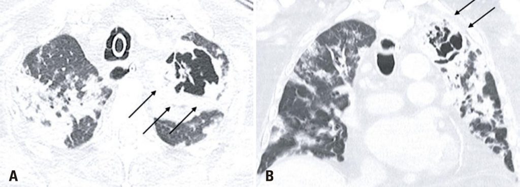
An 86-years-old male patient was admitted to the emergency department with a 1-day history of fever, dyspnea, and cough. After an assessment, there were no criteria that justified the patient’s hospitalization, therefore, he was dismissed and instructed to continue treatment at home. One week after the onset of symptoms, the patient returned with worsening dyspnea and persistent fever, which prompted his hospitalization. A chest computed tomography (CT) demonstrated typical coronavirus disease (COVID-19) findings(,) (), and the real-time polymerase chain reaction (rt-PCR) test confirmed the diagnosis. After 13 days of hospitalization, the patient experienced clinical worsening, and nosocomial pneumonia caused by Enterococcus faecalis was diagnosed (). The following CT 10 days after the diagnosis of the nosocomial infection showed multiple bilateral areas of ground-glass opacities, accompanied by septal thickening and consolidation areas, possibly related to COVID-19. There was also an excavated new lesion in the left upper lobe (). A new rt-PCR test was still positive for COVID-19 on the same day of this last chest CT scan.
[…]
3,956