Article

Oxidative Inactivation of SARS-CoV-2 on Photoactive AgNPs@TiO2 Ceramic Tiles

1
Department of Chemistry, University of Milan, Via Golgi 19, 20133 Milan, Italy
2
Department of Biomedical, Surgical and Dental Sciences, University of Milan, Via Carlo Pascal 36, 20133 Milan, Italy
3
Department of Pharmacological and Biomolecular Sciences, University of Milan, Via Carlo Pascal 36, 20133 Milan, Italy
4
Department of Biomedical Sciences for Health, University of Milan, Via Carlo Pascal 36, 20133 Milan, Italy
5
Department of Chemistry, University of Turin, Via Pietro Giuria 7, 10125 Turin, Italy
*
Authors to whom correspondence should be addressed.
Int. J. Mol. Sci. 2021, 22(16), 8836; https://doi.org/10.3390/ijms22168836
Submission received: 1 August 2021 / Revised: 13 August 2021 / Accepted: 15 August 2021 / Published: 17 August 2021
(This article belongs to the Special Issue Materials for Infectious Diseases)

Abstract

:
The current SARS-CoV-2 pandemic causes serious public health, social, and economic issues all over the globe. Surface transmission has been claimed as a possible SARS-CoV-2 infection route, especially in heavy contaminated environmental surfaces, including hospitals and crowded public places. Herein, we studied the deactivation of SARS-CoV-2 on photoactive AgNPs@TiO2 coated on industrial ceramic tiles under dark, UVA, and LED light irradiations. SARS-CoV-2 inactivation is effective under any light/dark conditions. The presence of AgNPs has an important key to limit the survival of SARS-CoV-2 in the dark; moreover, there is a synergistic action when TiO2 is decorated with Ag to enhance the virus photocatalytic inactivation even under LED. The radical oxidation was confirmed as the the central mechanism behind SARS-CoV-2 damage/inactivation by ESR analysis under LED light. Therefore, photoactive AgNPs@TiO2 ceramic tiles could be exploited to fight surface infections, especially during viral severe pandemics.

1. Introduction

In December 2019, an unknown SARS-CoV-2 virus was detected in the middle of China (Wuhan) [1], and on March 2020, the World Health Organization (WHO) declared it as a COVID-19 pandemic [2]. COVID-19 pathophysiology in most cases results in acute respiratory distress syndrome (ARDS) and gastrointestinal damage, and it also affects the nervous system, whereas elderly-aged people are more vulnerable to COVID-19 complications, especially those with a chronic critical illness (CI) [3,4]. To date, many infection cases and high daily mortality are still recording among the global populations, and the WHO worldwide recorded more than 208 million confirmed COVID-19 cases, including more than 4.3 million deaths in mid-August 2021.
COVID-19 was characterized by its highly person-to-person infection transmission via several routes [5,6]. Microdroplets can remain longer in the air, which elicits the risk of infections at up to 2 m from the infected person [7]. The indirect transmission through infected objects and environmental surfaces has also been considered if susceptible people touch contaminated objects/surfaces and then transfer the virus to themselves. This transmission might happen in highly viral contaminated places such as infected people’s rooms, clinics, and hospitals [5,8] but even in more common areas such as supermarkets, shops, gyms, restaurants, etc. The COVID-19 contamination in Wuhan (China) was found to be heavy in intensive care rooms, wherein the highly COVID-19 contamination was detected on floors and objects (bed, computer mice, trash cans, etc.) [9] and in infected patients’ hospital rooms [10,11]. International health bodies, including the WHO, ask for the implementation of social distancing, hand washing, continuous cleaning of objects and surfaces, and droplet precautions [12,13]. In May 2020, the WHO published a guidance report entitled Cleaning and disinfection of environmental surfaces in the context of COVID-19 [14], after confirming COVID-19 transmission through contaminated environmental surfaces [15]. In this report, the WHO suggested using chlorine-based disinfectants in particular for those situations in which the cleaning of surfaces using common disinfectants [16] is inconvenient in emergency cases, or even while some disinfectants are ineffective such as the common chlorhexidine digluconate disinfectant [17]. We have to remember that the over-use of disinfectant sprays and products might lead to severe health effects associated primarily with asthma and respiratory disease [18,19].
The lifetime of SARS-CoV-2 on different objects significantly depends on the nature of materials and environmental characteristics [20]. It was widely reported that SARS-CoV-2 stays alive mostly on smooth surfaces, such as windows, smooth ceramics, doorknobs, countertops, etc. Unlike other previous coronaviruses, some reports mentioned that SARS-CoV-2 can survive up to 21 days on environmental surfaces, which highly increases the chance of transmission [21,22].
In the present research, we suggest the employment of photocatalytic self-cleaning surfaces functionalized with silver-decorated TiO2 (AgNPs@TiO2 tiles) as an eco-technology in areas likely to be exposed to severe viral/bacterial infections such as clinics and hospitals to prevent the surface transmissions of microbial pathogens, including COVID-19. The photocatalytic concept is based on the surface coating of ceramic materials with photoactive compounds that, under light irradiation, produce highly oxidative radical oxygen species (ROSs). In turn, onto a photocatalytic surface, pollutants or pathogenic species undergo continuous degradation processes via ROSs [23,24]. In this specific case, the co-presence of Ag and TiO2 allows the final material to possess all the photocatalytic properties given by the photocatalytic coating along with the antibacterial/antiviral property already in the dark, thanks to the Ag action [25,26].
In this investigation, we tested the antiviral activity of industrially coated AgNPs@TiO2 tiles against the deactivation of SARS-CoV-2 in both dark and light conditions. The photocatalytic activity was already verified both on the bare powder and the industrially digitally printed ceramic tile, against different toxic air/water compounds such as organic dyes [27], drugs [28], and in abatement processes of NOx [29,30] and phenol [31]. Different bacteria strains were investigated, and both the complete degradation and the absence of the biofilm formation were also confirmed [26,32]. The crucial role of AgNPs@TiO2 in pathogenic inactivation is due to the excellent combination of AgNPs species, which is known for its natural antimicrobial activity [33,34], and the synergetic photocatalytic effects of TiO2 [35,36].

2. Results and Discussion

2.1. Ag-TiO2 Tile Characterization

The HR-TEM image in (Figure 1a) shows that TiO2 possesses the typical features of a micrometric titania system, with ordered and roundish particles (exhibiting diameter larger than 100 nm and typical anatase phase [28], on top of which Ag species are evident. The latter species could be found both in metallic form (Ag0, as indicated by the FFT analysis as shown in Figure S1) and Ag2+ species (in the form of Ag2O, as already reported in previously detailed research devoted to this topic [26,29]).
As for the morphology of the Ag@TiO2 particles on the surface tile, they have been characterized by HR-SEM investigation (Figure 1b). TiO2 particles exhibit sizes > 100 nm range (mean diameter), confirming thus the indications coming from HR-TEM observations (see above), with a quite uniform distribution of TiO2. EDX mappings relative to either Ti or Ag (Figure 1d,e) confirm this evidence, indicating a good distribution of Ti and Ag species on the surface of the engineered tile. Then, the tile surface was investigated by XPS to verify the composition of the external layer of the engineered surface: the analysis has been carried out both before and after the digital coating process, and survey spectra are reported in (Figure 1e). Characteristic peaks of Ti2p and Ag3d were detected for the AgNPs@TiO2 coated tile [37], as well as Si 2p and 2s peaks due to the silicate present in the ink formulation [38].

2.2. Antiviral Results

For comparison, antiviral experiments were performed on glass and AgNPs@TiO2 tile surfaces in dark and light irradiation (UVA or LED) with a starting viral load of 1.4 × 10⁵ PFU/cm2 (log10 5.146). The antiviral activity for SARS-CoV-2 was expressed in log10 reduction as described (see Section 2.1). Figure 2 shows comparatively the results of SARS-CoV-2 inactivation under different conditions. Detailed results are shown in Table S1.
In the dark, the inactivation of SARS-CoV-2 was more pronounced on the surface of AgNPs@TiO2 tile compared to the glass surface within 4 h. Many reports declared that the lifetime of SARS-CoV-2 is longer on a smooth surface such as glasses. The chance of SARS-CoV-2 survival depends on the interface interaction of SARS-CoV-2 droplets, contact angle humidity, and temperature [39]. Due to the smooth surface of glasses, the lifetime of droplets would be longer compared to ceramics materials. In addition, the Ag-rich tiles might prevent the survival or/and inactivate SARS-CoV-2 due to the antiviral effect of AgNPs [40]. Jeremiah et al. [41] studied the antiviral activity of AgNPs against SARS-CoV-2, and it was found that 10 nm diameter AgNPs showed excellent inhibition of extracellular SARS-CoV-2. On the glass surface under UVA, a 1.042 and 1.359 log10 reduction was observed within 4 and 7 h, respectively. Under these circumstances and compared with the experiment on glass in the dark, we may confirm that the intensity of UVA radiation at the range of 315–400 nm is not strong enough to deactivate SARS-CoV-2 effectively. Several reports investigated the inactivation of SARS-CoV-2 by direct deep UV irradiation [42,43,44]. Heilingloh et al. [45] reported that SARS-CoV-2 was very susceptible to UVC irradiation, while a low inactivation was found under UVA. UVC is the most common radiation used for the inactivation of viruses due to its germicidal effect wavelength peak, which fits with the absorption of nucleic acids [46,47].
In terms of Ag-TiO2 tile under UVA, the inactivation of the virus is much higher compared to that on the glass surface. Analysis of SARS-CoV-2 inactivation indicated that a 1.775 and 2.620 log10 reduction (corresponding to 98.3% and 99.7% viral reduction) was obtained at 4 and 7 h of contact time, respectively. To check the performance in the presence of higher UVA radiation, an experiment was carried out under UVA with an intensity of 0.25 mW/cm2 with a starting log10 PFU/cm2 of 3.079, and complete inactivation of SARS-CoV-2 was recorded within 7 h of irradiation.
To exclude any direct cytotoxicity of the surface wash solution on the host cells, a cytotoxicity assay was performed using broth recovered by the glass or Ag@TiO2 surface. As shown in the Supplementary Material Table S2, no cytotoxicity against VERO cells was observed.
In UV-exposed Ag@TiO2 tile, the inactivation of SARS-CoV-2 is due to the generation of reactive oxygen species (ROSs) due to the photoexcitation of Ag@TiO2 coated on the surface of the ceramic tile. The AgNPs and TiO2 heterojunction system in the presence of light irradiation can be very powerful for the generation of high yield of ROSs, e.g., OH radicals as already demonstrated in the E. Coli degradation in our previous study [26], in which the oxidative inactivation of E. Coli was investigated on the same samples (surface of Ag@TiO2 tiles) used in the present research. In that work, we observed that the light irradiation of the photocatalytic engineered surface led to shifting the interfacial potential due to (i) the surface stabilization and (ii) the photoproduction of ROSs on the active surface, bringing about the damage of E. Coli species. An oxidative synergistic effect resulting from the most photoproduced ROSs (OH, −•O2, and H2O2), together with the direct inactivation via the positive holes on the valence band of Ag@TiO2 are cooperatively responsible for the damage of microbial membranes. Long-lived H2O2 is known to be very powerful in terms of bacterial/viral inactivation because of its relative stability, which is requested for effective damage of microbial membrane [47,48,49], and this latter could be photocatalytically produced from the dimerization and reduction of OH and −•O2, respectively.
Some research groups using different photocatalysts, including TiO2, have reported the photocatalytic abatement of SARS-CoV-2 through oxidative damage [48,49,50]. Under LED light, the log10 reduction values were 1.323 and 2.210 within 7 h on glass and Ag@TiO2 tile, respectively, confirming the importance of both light irradiation and the presence of a photocatalytic surface to accelerate the viral inactivation.
The photocatalytic generation of OH species to confirm the radical oxidation of the SARS-CoV-2 was checked by ESR analysis under similar solar light with UV light cut-off (400 nm) using DMPO as a trapping agent: the relevant spectra are reported in Figure 3. Unlike bare TiO2, the pattern of the typical signals of DMPO-OH adduct [51] was notably detected in Ag@TiO2, confirming the high photonic synergism obtained in Ag-TiO2.
Based on the obtained results, two major mechanistic pathways could take place on the surface of Ag@TiO2 porcelain-grès tiles toward SARS-CoV-2 inactivation (Figure 4). In the dark, slow direct inactivation of SARS-CoV-2 can take place by AgNPs deposited on the surface. AgNPs can directly destroy the membrane of virus, and also, it is an excellent inhibitor against microbial growth. Under the light, fast deactivation of SARS-CoV-2 is a result of the radical oxidation which is produced continuously on the surface of photoactive tiles under UVA or LED. In real conditions, the yield of the spreading virus would be much lesser than the starting yield of SARS-CoV-2 tested in this study. Therefore, antiviral ceramics would be an excellent option mainly to prevent viral transmission, including SARS-CoV-2, in highly contaminated environments during the critical viral situation, especially in hospitals [52].

3. Materials and Methods

3.1. Fabrication of Ag-Decorated TiO2 Photoactive Tiles

Firstly, the Ag-decorated TiO2 powder was prepared by the impregnation method [25]. Commercial 1077-Kronos with particle size in the 110–130 nm range and surface area of 12 ± 2 m2g−1 was employed as a TiO2 photocatalyst source. AgNPs were synthesized starting from silver nitrate (AgNO3, ACS Reagent, Sigma-Aldrich, ≥99%), as Ag precursor, in the presence of KNO3 (ACS Reagent, Sigma-Aldrich ≥ 99.0%) and polyvinylpyrrolidone (PVP40, average mol·wt−1: 40,000, Sigma-Aldrich). AgNPs ratio in the Ag@TiO2 composite is 8%. The coating of tiles was industrially produced by digital printing (Projecta Engineering S.r.l., Fiorano M.se, Italy) on porcelain-grès tiles (IrisCeramica Group—Active R&D production site Castellarano, Italy) and stabilized in a kiln at 680 °C; details of this process were provided in previous works [26,53].

3.2. Antiviral Experiments and Calculations

3.2.1. Photocatalytic Antiviral Tests

Photocatalytic experiments were carried out in the dark and using a mercury UVA lamp (500W, Jelosil, Vomodrone, Italy) at 0.1 and 0.25 mW/m2 or a LED (Philips, Germany) at 1000 lux conditions, for 4 and 7 h. Experiments were carried out using glass plates for comparison purposes. Glass was properly chosen as the tile surfaces, which are glazed due to the presence of silicate in the coating formulation and its stabilization at 680 °C.

3.2.2. Viruses and Cells

SARS-CoV-2 was isolated from a nasal-pharyngeal swab positive for SARS-CoV-2. The complete nucleotide sequence of the SARS-CoV-2 isolated strain was deposited at Gen Bank, at NCBI (accession number: MT748758).
VERO (Monkey Kidney Epithelial Cells, clone E6, ATCC CRL-1586™) cells were maintained in DMEM medium (EuroClone, Pero, Italy) supplemented with 10% heat-inactivated fetal calf serum, 2 mM glutamine, 100 units/mL of penicillin, and 100 μg/mL of streptomycin (EuroClone, Pero, Italy).

3.2.3. Preparation of Test Specimens

Each sample (a flat square of (50 ± 2) mm × (50 ± 2) mm) was sterilized by immersion in ethanol 70%, to eliminate any bacterial contamination.

3.2.4. Test Procedure

ISO 18,061 protocol was chosen to test the samples with modifications. Both glass and Ag@TiO2 tile were inoculated with 0.2 mL of virus suspensions (1–5 × 10⁶ Plaque-Forming Unit (PFU)/mL), and the inoculum was covered with a 40 × 40 mm film and incubated for 4 or 7 h at room temperature in the dark or under the selected lighting system (LED or UVA).
At the end of the contact time, 20 mL of neutralizer SCDLP broth were added to the samples and plaque assay was performed, in 6-well plates, testing 4-fold serial dilutions of the recovered SCDLP broth in complete medium. Briefly, the cells monolayer was inoculated for 2 h, with 0.4 mL of the virus suspension, recovered in SCDLP broth; each dilution was tested in duplicate. Then, the inoculum was removed, the cells were washed with PBS, covered with 0.3% agarose dissolved in cell medium, and incubated for 48 h at 37 °C, 5% CO2. Cells were fixed with 4% formaldehyde solution (Sigma-Aldrich) and, after agarose removal, stained with methylene blue (Sigma-Aldrich). Plaques were counted, and the infectivity titer of the virus was expressed as PFU/cm2. The antiviral activity for SARS-CoV-2 was expressed in log10 reduction (log10 PFU/cm2 at Time 0 (t0)-log10 PFU/cm2 at the subsequent time points).
At T = 0, immediately after virus inoculum, 20 mL of neutralizer SCDLP broth were added to 3 glass samples, and the residual virus infectivity was revealed by plaque assay.

3.2.5. Cytotoxicity and Cell Sensitivity to Virus

For the cytotoxicity assay, cells were seeded into 96-well plates at a concentration of 1.3×10⁴ cells/well. Twenty mL of neutralizer SCDLP broth were added to 3 glass and 3 photoactive samples, and immediately, 0.1 mL was recovered and added to the cells in triplicate. After 2 h of incubation, the SCDLP broth was replaced with a complete medium, and cells were incubated for 48 h at 37 °C in 5% CO2. At the end of incubation, cell viability was measured by MTT (3-[4.5-dimethylthiazol-2-yl]-2.5-diphenyltetrazolium bromide) assay. Twenty µL of MTT solution (5 mg/mL) was added to each well for 3 h. Then, the plates were centrifuged, the supernatants discarded, and the resulting pellets dissolved in 100 µL of lysing buffer consisting of 20% (w/v) of a solution of SDS (Sigma-Aldrich), 40% of N,N-dimethylformamide (Sigma-Aldrich)) in H2O. The absorbance was measured spectrophotometrically at a test wavelength of 550 nm and a reference wavelength of 650 nm, using a Synergy 4 microplate reader (Biotek, GE).

3.3. Materials Characterization
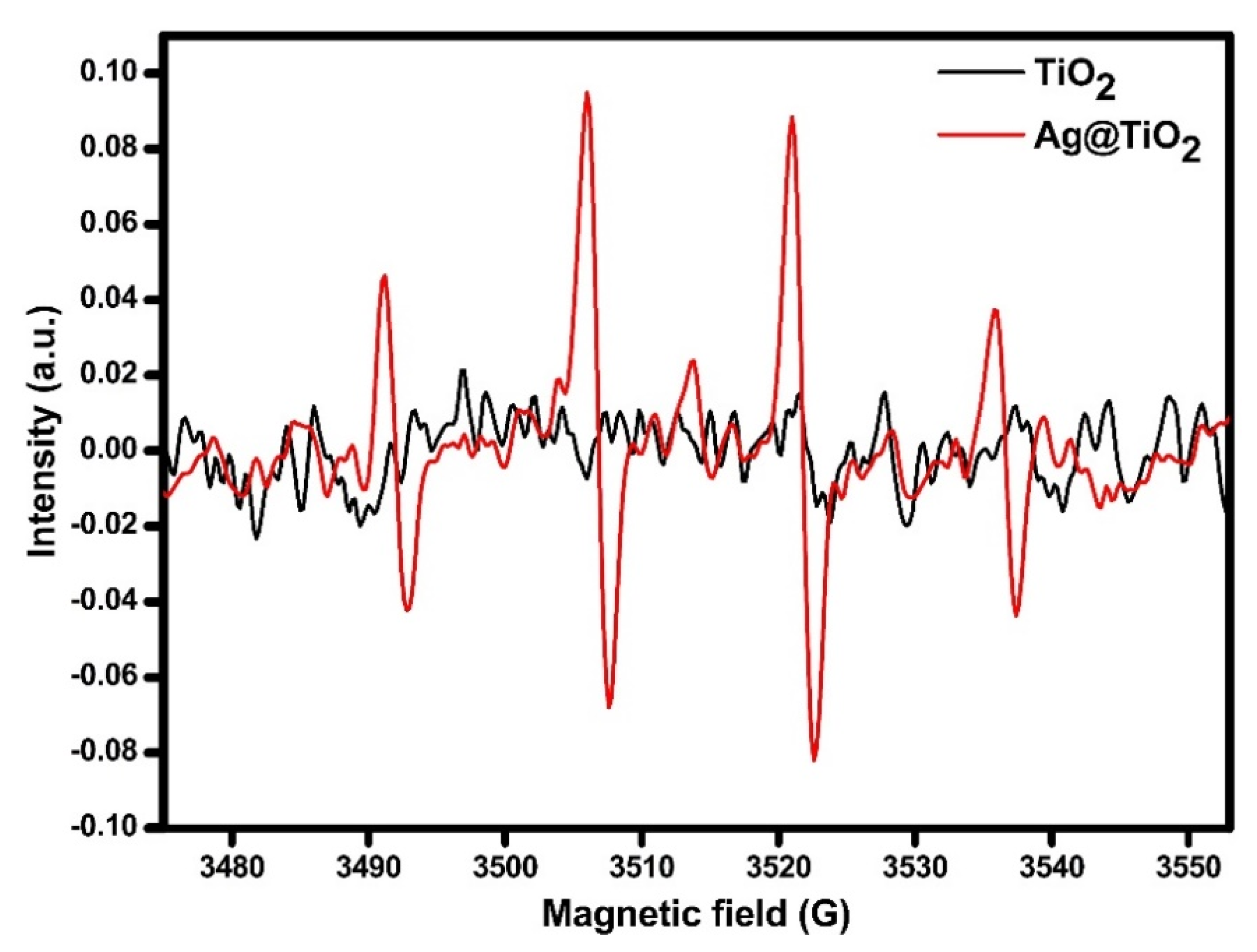
3.3.1. DMPO-OH ESR Analysis

ESR analysis to check the photocatalytic generation of OH on powdered Kronos 1077 TiO2 and the same sample decorated (Ag-TiO2) was carried out by using as the irradiating source a solar box (Co. Fo. Megra, Milan, Italy) equipped with a 1500 W Xenon lamp and cut-off filters for wavelengths below 340 nm or 400 nm. Then, 3 mL of a sample suspension (prepared to introduce 100 mg of sample in 100 mL of pure water) were introduced in a quartz cell and irradiated under stirring for 20 min in the presence of 5,5-dimethyl-1-pyrroline-N-oxide (DMPO, 17 mM). ESR spectra were recorded at room temperature using an X-band Bruker-EMX spectrometer equipped with a cylindrical cavity operating at 100 kHz field modulation. Experimental parameters were as follows: microwave frequency 9.86 GHz; microwave power 2.7 mW; modulation amplitude 2 Gauss; conversion time 30.68 ms.

3.3.2. HR-TEM Characterization

HR-TEM images have been obtained employing a Jeol JEM 3010-UHR (Japan) microscope equipped with LaB6 filament (potential acceleration 300 kV). Images were digitally acquired using an Ultrascan 1000 camera and processed with Gatan Digital Micrograph program version 3.11.1. Before the analysis, samples were dry dispersed onto Cu grids coated with lacey carbon film.

3.3.3. HR-SEM Characterization

A Field Emission Electron Scanning Microscope (FE-SEM) LEO 1525 ZEISS (Germany) was used to determine the photocatalyst distribution at the ceramic surface. Samples were deposited on conductive carbon adhesive tape and metalized with chromium.

3.3.4. X-ray Photoelectron Spectroscopy

An M-probe apparatus (XPS–M-Probe, Surface Science Instruments, USA) recorded the XPS spectra. The instrument is equipped with a monochromatic AlKα anode and a C1s peak at 284.6 eV was used as internal calibration [54]. The energy scale was calibrated with reference to the 4f7/2 level of a freshly evaporated gold sample, which was taken as 84.00 eV and with reference to the 2p3/2 level of copper taken as 932.47 ±0.10 eV and to the 3s level of copper (122.39 ± 0.15 eV), respectively. An electron gun at 7 eV was used for the analyses of insulating samples.

4. Conclusions

In the present report, we showed the antiviral activity of AgNPs-decorated TiO2 based photoactive ceramic tiles toward SARS-CoV-2 under dark, UVA, and LED light irradiations. Compared to control experiments on a glass surface, Ag@TiO2 photoactive ceramic tiles showed faster SARS-CoV-2 inactivation. In dark conditions, the inactivation of SARS-CoV-2 is several times faster on Ag-TiO2 ceramic tile than on glass. In addition, UVA irradiation significantly boosts the inactivation of SARS-CoV-2 on Ag@TiO2 ceramic tile via radical oxidation, which was confirmed by ESR analysis. Total SARS-CoV-2 inactivation with starting Log PFU/cm2 = 3.079 was found using UVA intensity of 0.25 mW/cm2. Under UVA at a lower intensity of 0.1 mW/cm2, high inactivation rates were recorded as well. The radical oxidative inactivation of SARS-CoV-2 was also found under LED indoor light irradiation. The results of this investigation showed the potential of self-cleaning photoactive materials toward viral inactivation. Exploitation and extending this sustainable technology in heavy contaminated environmental surfaces may help to reduce viral surface transmission, especially during a strong viral pandemic such as the case of SARS-CoV-2.

Supplementary Materials

The following are available online at https://www.mdpi.com/article/10.3390/ijms22168836/s1.

Author Contributions

S.D. (Sarah D’Alessandro) and S.P.; investigation, N.B. and E.F.; resources, C.L.B.; data curation, S.D. (Serena Delbue); formal analysis, G.C., R.D. and E.L.; writing—original draft preparation, R.D. and N.B.; writing—review and editing, R.D. and C.L.B.; supervision, C.L.B. All authors have read and agreed to the published version of the manuscript.

Funding

The University of Milan, APC fund, is acknowledged by the authors for financial support.

Institutional Review Board Statement

Not applicable.

Informed Consent Statement

Not applicable.

Data Availability Statement

Not applicable.

Acknowledgments

The authors would like to thank Iris Ceramica Group for providing the photocatalytic porcelain-grès samples from the Calacatta SL 300 × 150 Active Surfaces series.

Conflicts of Interest

The authors declare no conflict of interest.

References

  1. World Health Organization. Novel Coronavirus (2019-nCoV): Situation Report, 3; World Health Organization: Geneva, Switzerland, 2020. [Google Scholar]
  2. World Health Organization. WHO Director-General’s Opening Remarks at the Media Briefing on COVID-19—11 March 2020; World Health Organization: Geneva, Switzerland, 2020. [Google Scholar]
  3. Pollard, C.A.; Morran, M.P.; Nestor-Kalinoski, A.L. The COVID-19 pandemic: A global health crisis. Physiol. Genom. 2020, 52, 549–557. [Google Scholar] [CrossRef]
  4. Parasher, A. COVID-19: Current understanding of its Pathophysiology, Clinical presentation and Treatment. Postgrad. Med. J. 2021, 97, 312–320. [Google Scholar] [CrossRef]
  5. Jin, H.; Hong, C.; Chen, S.; Zhou, Y.; Wang, Y.; Mao, L.; Li, Y.; He, Q.; Li, M.; Su, Y.; et al. Consensus for prevention and management of coronavirus disease 2019 (COVID-19) for neurologists. Stroke Vasc. Neurol. 2020, 5, 146–151. [Google Scholar] [CrossRef] [Green Version]
  6. Harrison, A.G.; Lin, T.; Wang, P. Mechanisms of SARS-CoV-2 transmission and pathogenesis. Trends Immunol. 2020, 41, 1100–1115. [Google Scholar] [CrossRef]
  7. Chagla, Z.; Hota, S.; Khan, S.; Mertz, D. Re: It Is Time to Address Airborne Transmission of COVID-19. Clin. Infect. Dis. 2020. [Google Scholar] [CrossRef]
  8. Lei, H.; Xu, X.; Xiao, S.; Wu, X.; Shu, Y. Household transmission of COVID-19—A systematic review and meta-analysis. J. Infect. 2020, 81, 979–997. [Google Scholar] [CrossRef]
  9. Guo, Z.-D.; Wang, Z.-Y.; Zhang, S.-F.; Li, X.; Li, L.; Li, C.; Cui, Y.; Fu, R.-B.; Dong, Y.-Z.; Chi, X.-Y.; et al. Aerosol and surface distribution of severe acute respiratory syndrome coronavirus 2 in hospital wards, Wuhan, China, 2020. Emerg. Infect. Dis. 2020, 26, 1586–1591. [Google Scholar] [CrossRef]
  10. Ong, S.W.X.; Tan, Y.K.; Chia, P.Y.; Lee, T.H.; Ng, O.T.; Wong, M.S.Y.; Marimuthu, K. Air, surface environmental, and personal protective equipment contamination by severe acute respiratory syndrome coronavirus 2 (SARS-CoV-2) from a symptomatic patient. JAMA 2020, 323, 1610–1612. [Google Scholar] [CrossRef] [Green Version]
  11. Yung, C.F.; Kam, K.-Q.; Wong, M.S.; Maiwald, M.; Tan, Y.K.; Tan, B.H.; Thoon, K.C. Environment and personal protective equipment tests for SARS-CoV-2 in the isolation room of an infant with infection. Ann. Intern. Med. 2020, 173, 240–242. [Google Scholar] [CrossRef] [Green Version]
  12. McIntosh, K.; Hirsch, M.; Bloom, A. Coronavirus disease 2019 (COVID-19): Epidemiology, virology, and prevention. Lancet. Infect. Dis. 2020, 1, 2019–2020. [Google Scholar]
  13. Arora, V.M.; Chivu, M.; Schram, A.; Meltzer, D. Implementing physical distancing in the hospital: A key strategy to prevent nosocomial transmission of COVID-19. J. Hosp. Med. 2020, 15, 290–291. [Google Scholar] [CrossRef] [Green Version]
  14. World Health Organization. Cleaning and Disinfection of Environmental Surfaces in the Context of COVID-19: Interim Guidance, 15 May 2020; World Health Organization: Geneva, Switzerland, 2020. [Google Scholar]
  15. Cheng, V.C.; Wong, S.-C.; Chen, J.H.; Yip, C.C.; Chuang, V.W.; Tsang, O.T.Y.; Sridhar, S.; Chan, J.F.W.; Ho, P.-L.; Yuen, K.-Y. Escalating infection control response to the rapidly evolving epidemiology of the coronavirus disease 2019 (COVID-19) due to SARS-CoV-2 in Hong Kong. Infect. Control Hosp. Epidemiol. 2020, 41, 493–498. [Google Scholar] [CrossRef] [Green Version]
  16. Rutala, W.A.; Weber, D.J. Disinfectants used for environmental disinfection and new room decontamination technology. Am. J. Infect. Control 2013, 41, S36–S41. [Google Scholar] [CrossRef]
  17. Assis, M.S.D.; Araújo, R.A.D.A.M.; Lopes, A.M.M. Safety alert for hospital environments and health professional: Chlorhexidine is ineffective for coronavirus. Rev. Assoc. Méd. Bras. 2020, 66, 124–129. [Google Scholar] [CrossRef]
  18. Zock, J.-P.; Plana, E.; Jarvis, D.; Antó, J.M.; Kromhout, H.; Kennedy, S.M.; Künzli, N.; Villani, S.; Olivieri, M.; Torén, K.; et al. The use of household cleaning sprays and adult asthma: An international longitudinal study. Am. J. Respir. Crit. Care Med. 2007, 176, 735–741. [Google Scholar] [CrossRef] [Green Version]
  19. Casey, M.L.; Hawley, B.; Edwards, N.; Cox-Ganser, J.M.; Cummings, K.J. Health problems and disinfectant product exposure among staff at a large multispecialty hospital. Am. J. Infect. Control 2017, 45, 1133–1138. [Google Scholar] [CrossRef] [PubMed]
  20. Joonaki, E.; Hassanpouryouzband, A.; Heldt, C.L.; Areo, O. Surface chemistry can unlock drivers of surface stability of SARS-CoV-2 in variety of environmental conditions. Chem 2020, 6, 2135–2146. [Google Scholar] [CrossRef]
  21. Marzoli, F.; Bortolami, A.; Pezzuto, A.; Mazzetto, E.; Piro, R.; Terregino, C.; Bonfante, F.; Belluco, S. A systematic review of human coronaviruses survival on environmental surfaces. Sci. Total Environ. 2021, 778, 146191. [Google Scholar] [CrossRef]
  22. Kasloff, S.B.; Leung, A.; Strong, J.E.; Funk, D.; Cutts, T. Stability of SARS-CoV-2 on critical personal protective equipment. Sci. Rep. 2021, 11, 1–7. [Google Scholar] [CrossRef] [PubMed]
  23. Kamegawa, T.; Shimizu, Y.; Yamashita, H. Superhydrophobic surfaces with photocatalytic self-cleaning properties by nanocomposite coating of TiO2 and polytetrafluoroethylene. Adv. Mater. 2012, 24, 3697–3700. [Google Scholar] [CrossRef] [PubMed]
  24. Tung, W.S.; Daoud, W.A. Self-cleaning fibers via nanotechnology: A virtual reality. J. Mater. Chem. 2011, 21, 7858–7869. [Google Scholar] [CrossRef]
  25. Minozzi, F.; Bianchi, C.L.; Pellini, R.; Capucci, V. Photocatalytic particles and Process for the Production. Thereof. Patent EP3277634B1, 5 February 2020. [Google Scholar]
  26. Bianchi, C.L.; Cerrato, G.; Bresolin, B.M.; Djellabi, R.; Rtimi, S. Digitally Printed AgNPs Doped TiO2 on Commercial Porcelain-Grès Tiles: Synergistic Effects and Continuous Photocatalytic Antibacterial Activity. Surfaces 2020, 3, 11–25. [Google Scholar] [CrossRef] [Green Version]
  27. Bianchi, C.L.; Colombo, E.; Gatto, S.; Stucchi, M.; Cerrato, G.; Morandi, S.; Capucci, V. Photocatalytic degradation of dyes in water with micro-sized TiO2 as powder or coated on porcelain-grès tiles. J. Photochem. Photobiol. A Chem. 2014, 280, 27–31. [Google Scholar] [CrossRef]
  28. Bianchi, C.L.; Sacchi, B.; Capelli, S.; Pirola, C.; Cerrato, G.; Morandi, S.; Capucci, V. Micro-sized TiO2 as photoactive catalyst coated on industrial porcelain grès tiles to photodegrade drugs in water. Environ. Sci. Pollut. Res. 2018, 25, 20348–20353. [Google Scholar] [CrossRef]
  29. Cerrato, G.; Galli, F.; Boffito, D.C.; Operti, L.; Bianchi, C.L. Correlation preparation parameters/activity for microTiO2 decorated with SilverNPs for NOx photodegradation under LED light. Appl. Catal. B Environ. 2019, 253, 218–225. [Google Scholar] [CrossRef] [Green Version]
  30. Bianchi, C.; Pirola, C.; Galli, F.; Vitali, S.; Minguzzi, A.; Stucchi, M.; Manenti, F.; Capucci, V. NO x degradation in a continuous large-scale reactor using full-size industrial photocatalytic tiles. Catal. Sci. Technol. 2016, 6, 2261–2267. [Google Scholar] [CrossRef] [Green Version]
  31. Bianchi, C.L.; Stucchi, M.; Pirola, C.; Cerrato, G.; Sacchi, B.; Vitali, S.; Di Michele, A.; Capucci, V. Micro-sized TiO2 catalyst in powder form and as coating on porcelain grès tile for the photodegradation of phenol as model pollutant for water phase. Adv. Mater. Sci. 2017, 2. [Google Scholar] [CrossRef] [Green Version]
  32. Bianchi, C.L.; Pirola, C.; Stucchi, M.; Sacchi, B.; Cerrato, G.; Morandi, S.; Di Michele, A.C.A.; Capucci, V. A New Frontier of Photocatalysis Employing Micro-Sized TiO2: Air/Water Pollution Abatement and Self-Cleaning/Antibacterial Applications; IntechOpen: London, UK, 2016; pp. 635–666. [Google Scholar]
  33. Kim, J.S.; Kuk, E.; Yu, K.N.; Kim, J.-H.; Park, S.; Lee, H.J.; Kim, S.H.; Park, Y.K.; Park, Y.H.; Hwang, C.-Y.; et al. Antimicrobial effects of silver nanoparticles. Nanomed. Nanotechnol. Biol. Med. 2007, 3, 95–101. [Google Scholar] [CrossRef] [PubMed]
  34. Russell, A.; Hugo, W. 7 Antimicrobial Activity and Action of Silver. Prog. Med. Chem. 1994, 31, 351–370. [Google Scholar] [CrossRef] [PubMed]
  35. Wu, N. Plasmonic metal–semiconductor photocatalysts and photoelectrochemical cells: A review. Nanoscale 2018, 10, 2679–2696. [Google Scholar] [CrossRef] [PubMed]
  36. Hong, J.W.; Wi, D.H.; Lee, S.-U.; Han, S.W. Metal–semiconductor heteronanocrystals with desired configurations for plasmonic photocatalysis. J. Am. Chem. Soc. 2016, 138, 15766–15773. [Google Scholar] [CrossRef] [PubMed]
  37. Chen, H.; Huang, M.; Wang, Z.; Gao, P.; Cai, T.; Song, J.; Zhang, Y.; Meng, L. Enhancing rejection performance of tetracycline resistance genes by a TiO2/AgNPs-modified nanofiber forward osmosis membrane. Chem. Eng. J. 2020, 382, 123052. [Google Scholar] [CrossRef]
  38. Cañón, J.; Teplyakov, A.V. XPS characterization of cobalt impregnated SiO2 and γ-Al2O3. Surf. Interface Anal. 2021, 53, 475–481. [Google Scholar] [CrossRef]
  39. Bhardwaj, R.; Agrawal, A. Likelihood of survival of coronavirus in a respiratory droplet deposited on a solid surface. Phys. Fluids 2020, 32, 061704. [Google Scholar] [CrossRef]
  40. Galdiero, S.; Falanga, A.; Vitiello, M.; Cantisani, M.; Marra, V.; Galdiero, M. Silver nanoparticles as potential antiviral agents. Molecules 2011, 16, 8894–8918. [Google Scholar] [CrossRef] [Green Version]
  41. Jeremiah, S.S.; Miyakawa, K.; Morita, T.; Yamaoka, Y.; Ryo, A. Potent antiviral effect of silver nanoparticles on SARS-CoV-2. Biochem. Biophys. Res. Commun. 2020, 533, 195–200. [Google Scholar] [CrossRef]
  42. Inagaki, H.; Saito, A.; Sugiyama, H.; Okabayashi, T.; Fujimoto, S. Rapid inactivation of SARS-CoV-2 with deep-UV LED irradiation. Emerg. Microbes Infect. 2020, 9, 1744–1747. [Google Scholar] [CrossRef]
  43. Minamikawa, T.; Koma, T.; Suzuki, A.; Mizuno, T.; Nagamatsu, K.; Arimochi, H.; Tsuchiya, K.; Matsuoka, K.; Yasui, T.; Yasutomo, K.; et al. Quantitative evaluation of SARS-CoV-2 inactivation using a deep ultraviolet light-emitting diode. Sci. Rep. 2021, 11, 1–9. [Google Scholar]
  44. Gidari, A.; Sabbatini, S.; Bastianelli, S.; Pierucci, S.; Busti, C.; Bartolini, D.; Stabile, A.M.; Monari, C.; Galli, F.; Rende, M.; et al. SARS-CoV-2 Survival on Surfaces and the Effect of UV-C Light. Viruses 2021, 13, 408. [Google Scholar] [CrossRef]
  45. Heilingloh, C.S.; Aufderhorst, U.W.; Schipper, L.; Dittmer, U.; Witzke, O.; Yang, D.; Zheng, X.; Sutter, K.; Trilling, M.; Alt, M.; et al. Susceptibility of SARS-CoV-2 to UV irradiation. Am. J. Infect. Control 2020, 48, 1273–1275. [Google Scholar] [CrossRef] [PubMed]
  46. Seyer, A.; Sanlidag, T. Solar ultraviolet radiation sensitivity of SARS-CoV-2. Lancet Microbe 2020, 1, e8–e9. [Google Scholar] [CrossRef]
  47. Bono, N.; Ponti, F.; Punta, C.; Candiani, G. Effect of UV Irradiation and TiO2-Photocatalysis on Airborne Bacteria and Viruses: An Overview. Materials 2021, 14, 1075. [Google Scholar] [CrossRef] [PubMed]
  48. Horváth, E.; Rossi, L.; Mercier, C.; Lehmann, C.; Sienkiewicz, A.; Forró, L. Photocatalytic Nanowires-Based Air Filter: Towards Reusable Protective Masks. Adv. Funct. Mater. 2020, 30, 2004615. [Google Scholar] [CrossRef]
  49. Khaiboullina, S.; Uppal, T.; Dhabarde, N.; Subramanian, V.R.; Verma, S.C. Inactivation of Human Coronavirus by Titania Nanoparticle Coatings and UVC Radiation: Throwing Light on SARS-CoV-2. Viruses 2021, 13, 19. [Google Scholar] [CrossRef]
  50. Weiss, C.; Carriere, M.; Fusco, L.; Capua, I.; Regla-Nava, J.A.; Pasquali, M.; Scott, J.A.; Vitale, F.; Unal, M.A.; Mattevi, C.; et al. Toward nanotechnology-enabled approaches against the COVID-19 pandemic. ACS Nano 2020, 14, 6383–6406. [Google Scholar] [CrossRef]
  51. García-Negueroles, P.; García-Ballesteros, S.; Amat, A.M.; Laurenti, E.; Arques, A.; Santos-Juanes, L. Unveiling the Dependence between Hydroxyl Radical Generation and Performance of Fenton Systems with Complexed Iron. ACS Omega 2019, 4, 21698–21703. [Google Scholar] [CrossRef]
  52. Imani, S.M.; Ladouceur, L.; Marshall, T.; Maclachlan, R.; Soleymani, L.; Didar, T.F. Antimicrobial Nanomaterials and Coatings: Current Mechanisms and Future Perspectives to Control the Spread of Viruses Including SARS-CoV-2. ACS Nano 2020, 14, 12341–12369. [Google Scholar] [CrossRef]
  53. Bianchi, C.L.; Cerrato, G.; Pirola, C.; Galli, F.; Capucci, V. Photocatalytic porcelain grés large slabs digitally coated with AgNPs-TiO2. Environ. Sci. Pollut. Res. 2019, 26, 36117–36123. [Google Scholar] [CrossRef] [PubMed]
  54. Lefebvre, J.; Galli, F.; Bianchi, C.L.; Patience, G.-S.; Boffito, D.C. Experimental Methods in Chemical Engineering: X-ray Photoelectron Spectroscopy—XPS. Can. J. Chem. Eng. 2019, 97, 2588–2593. [Google Scholar] [CrossRef] [Green Version]
Figure 1. (a) HR-TEM images of Ag@TiO2 powder. (b) SEM images of 8% Ag-decorated TiO2 particles. (c,d) EDX mapping of Ti and Ag species, respectively, on the photocatalytic porcelain-grès tile. (e) XPS survey spectra of uncoated and Ag@TiO2 coated tiles.
Ijms 22 08836 g001
Figure 2. (a) Inactivation of SARS-CoV-2 under different conditions (dark, LED, UVA: 0.1 mW/cm) after 4 and 7 h. (b) Selected results under different dark/lighting conditions (LED and UVA: 0.1 and 0.25 mW/cm) after 7 h.
Ijms 22 08836 g002
Figure 3. ESR analysis using DMPO as a trapping agent under similar solar light with UV light cut-off (>400 nm).
Ijms 22 08836 g003
Figure 4. Mechanistic pathways of SARS-CoV-2 inactivation on the surface of Ag-decorated TiO2 porcelain-grès tiles with light irradiations (UVA or LED) or in the dark.
Ijms 22 08836 g004
Publisher’s Note: MDPI stays neutral with regard to jurisdictional claims in published maps and institutional affiliations.

